Vagus Nerve: Unlocking the Healing Potential

Vagus Nerve: 10th Cranial Nerve in the Body

Overview

Did you know there are 12 cranial nerves in your body?

They work in pairs to establish connections between your brain and different parts of your body, like your head, neck, and torso.

Some of these nerves carry sensory information, relaying details about smells, sights, tastes, and sounds to your brain. These nerves are responsible for your senses.

On the other hand, certain cranial nerves control the movement of muscles and the function of glands. These nerves are in charge of motor functions.

The vagus nerve is responsible for carrying sensory and motor signals between the brain and the body, playing a crucial role in regulating many bodily functions.

Let’s explore more about it:

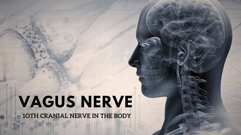
What is Vagus Nerve?

The vagus nerve, also known as the tenth cranial nerve or CN X, is one of the major nerves in the human body. It is a long and complex nerve that extends from the brainstem to various organs in the body, including the heart, lungs, stomach, and intestines.

The sensory nerve plays a big role in keeping our gut healthy. It controls the contraction of muscles in our intestines and sends messages to our brain and other important organs about what’s happening in our gut. This is known as the Brain-Gut Axis, and it helps to manage inflammation.

The vagus nerve, by regulating our breathing and heart rate, has a direct effect on how we respond to stress and how well we can relax.

The classification of cranial nerves uses Roman numerals to indicate their location.

Take a look at the diagram below to visualize the position of the vagus nerve.

The Position of the Vagus Nerve

When does the Vagus Nerve Develop?

The development of the vagus nerve occurs after birth. During the first year of life, an infant’s nervous system undergoes significant changes.

Initially, in the third trimester of pregnancy, the vagus nerve focuses on crucial functions like breathing, feeding, and regulating body temperature to adapt to life outside the womb.

As the infant grows, the vagus nerve continues to develop, allowing them to interact with their surroundings.

It enables physiological regulation of essential life functions, including breathing, eating, and heart rate.

This development throughout the first year enables infants to engage with objects, people, and their environment more actively.

What does the Vagus Nerve Control?

Vagus Nerve Controls our Brain

The vagus nerve, also known as the pneumogastric nerve, affects several important functions in the body, including:

  1. Digestion: It plays a role in regulating digestive processes, such as the release of enzymes and the movement of food through the digestive system.

  2. Heart rate: It helps regulate heart rate, promoting a balanced and healthy rhythm.

  3. Breathing: It influences breathing patterns and helps control the rate and depth of respiration.

  4. Cardiovascular activity: The vagus nerve contributes to the regulation of blood pressure and cardiovascular functions.

  5. Reflex actions: It is involved in reflex actions like coughing, sneezing, swallowing, and vomiting.

The vagus nerve is part of the autonomic nervous system, which manages involuntary actions in the body, including breathing and digestion.

How the Vagus Nerve Is Central to Infant Emotional Development

Vagus nerve emotional development in Infants

The vagus nerve is vital for infant emotional development. It regulates the autonomic nervous system, shaping emotional responses.

It helps establish bonding, promotes self-regulation, and influences long-term emotional well-being.

Higher vagal tone is linked to better emotional regulation and secure attachment, while lower tone may lead to difficulties.

Nurturing positive experiences and supportive caregiving are crucial for healthy vagal functioning and emotional resilience in infants.

Effects on Emotional Development

As mammals, we are unable to take care of ourselves independently, relying on others for a safe environment.

Our emotional development and the strength of our vagus nerve are greatly influenced by the care we receive from others.

When our basic needs, as well as our social and emotional needs, are met in a loving environment, it contributes to the development of a strong or high vagal tone.

This positive foundation facilitates a smooth transition from dependency on caregivers to becoming well-adjusted, independent individuals in adolescence and adulthood.

Problems with the Vagus Nerve

Vasovagal Syncope | Vagus Nerve |

When the vagus nerve is damaged, it can lead to various symptoms due to its extensive reach and impact on multiple areas of the body.

Some potential symptoms of vagus nerve damage include:

  1. Difficulty speaking or changes in voice.
  2. Difficulty swallowing or loss of the gag reflex.
  3. Low blood pressure or fluctuations in heart rate (either slow or fast).
  4. Digestive issues, such as changes in the digestive process, nausea, vomiting, abdominal bloating, or pain.
  5. Depression and anxiety in individuals with breathing problems or heart disease.

The specific symptoms experienced depend on the location of the nerve damage.

Heart Rate Issues

Vagal nerve dysfunction can result in both slow (bradycardia) and fast (tachycardia) heart rates, depending on the type of dysfunction.

Overactivity of the vagus nerve can lead to a slow heart rate, while inadequate activity can cause a fast heart rate.

In some cases, vagal nerve maneuvers may be used to stimulate more vagal nerve activity and slow down the heart rate.

Gastroparesis

Damage to the vagus nerve can contribute to a condition called gastroparesis, which affects the involuntary contractions of the digestive system and impairs stomach emptying.

Gastroparesis can sometimes occur after a vagotomy procedure, which involves the removal of all or part of the vagus nerve.

Vasovagal Syncope

The vagus nerve stimulates certain heart muscles involved in slowing down the heart rate. If the vagus nerve overreacts, it can cause a sudden drop in heart rate and blood pressure, resulting in fainting episodes known as vasovagal syncope.

Triggers for this condition can include pregnancy, emotional stress, pain, or may occur without a clear cause. Alongside fainting, symptoms may include

  • warmth
  • nausea
  • tunnel vision
  • ringing in the ears
  • excessive sweating
  • low blood pressure
  • slow or irregular heartbeat.

If experiencing fainting, it’s advisable to consult a doctor to rule out serious underlying causes.

Preventive measures may include staying well-hydrated or avoiding rapid changes in posture.

Vagus Nerve Stimulation (VNS) 

Vagus Nerve Stimulation

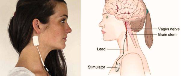
VNS is a treatment method that involves using electrical impulses to stimulate the vagus nerve. It is primarily used for cases of epilepsy and depression that do not respond to other treatments.

Researchers believe that the vagus nerve may play a role in linking depression, metabolic diseases, and heart disease. If ongoing research confirms this connection, VNS could potentially offer benefits for a variety of health issues.

During the VNS procedure, a device is typically implanted beneath the skin of the chest, and a wire connects it to the left vagus nerve. Once activated, the device sends signals through the vagus nerve to the brainstem, relaying information to the brain.

A neurologist usually programs the device, but individuals often receive a handheld magnet that allows them to control the device themselves.

It is believed that VNS holds potential for treating other conditions in the future, such as

  • Multiple sclerosis
  • Posterior autoimmune uveitis
  • Alzheimer’s disease
  • cluster headaches

However, further research is required to explore the effectiveness of VNS for these conditions.

Ongoing Research and Considerations

Researchers are actively investigating the vagus nerve as it holds potential clues to various health issues.

Here are some areas of interest:

  1. Mental health and well-being: There is interest in exploring different methods of stimulating the vagus nerve to improve mental well-being and manage depression. Techniques such as diaphragmatic breathing, cold water immersion, transcutaneous VNS devices, biofeedback, and yoga have been explored. However, the safety and effectiveness of these methods for everyone are not yet fully established, and it is important to consult a doctor before trying new treatments.

  2. Aging and vagal response: Research suggests that vagal response decreases with age. Individuals with a strong vagal tone may find it easier to relax after stressful events, exhibit better management of inflammation and gut issues, and potentially have improved blood sugar regulation. However, further research is needed to validate these findings.

  3. Rheumatoid arthritis (RA): There is some evidence that VNS may help reduce the severity of RA, an inflammatory condition affecting the joints. A study in 2021 found that individuals using a VNS device for 12 weeks experienced meaningful reductions in inflammation markers. However, more research is required to determine the safety and efficacy of VNS in managing Rheumatoid arthritis across a broader population.

It is essential to keep in mind that while these areas show promise, further research is necessary to establish the effectiveness, safety, and applicability of VNS and related techniques for specific conditions and individuals.

Consulting with a healthcare professional is crucial before attempting any new treatment strategy.

5 Ways to Activate The Vagus Nerve

Here are five effective ways to activate the vagus nerve:

  1. Deep Breathing: Engage in slow, deep breaths by inhaling deeply through your nose and exhaling slowly through your mouth. This type of diaphragmatic breathing stimulates the vagus nerve and promotes relaxation and stress reduction.

  2. Cold Exposure: Brief exposure to cold temperatures, such as taking cold showers or splashing cold water on your face, can activate the vagus nerve. The shock of cold triggers a natural physiological response that stimulates the nerve and enhances its function.

  3. Singing or Chanting: Vocal exercises like singing or chanting involve controlled breath and vibration, which stimulate the vagus nerve. This practice can help regulate your heart rate and promote a sense of calm and well-being.

  4. Meditation or Mindfulness: Engaging in meditation or mindfulness practices has been shown to activate the vagus nerve. Focusing on the present moment, deep breathing, and cultivating a state of relaxation can enhance vagal tone and overall well-being.

  5. Yoga or Tai Chi: These mind-body practices combine deep breathing, gentle movements, and relaxation techniques, all of which can activate the vagus nerve. Yoga poses, such as backbends or inversions, are particularly effective in stimulating the nerve.

It’s important to note that the effectiveness of these methods may vary from person to person, and it’s always a good idea to consult with a healthcare professional before trying new techniques, especially if you have any underlying health conditions.

Take Care of Yourself!

Share:

Facebook
WhatsApp
Aviral
Reviewed By : Dr. Aviral Vatsa

Social Media

Most Popular

Get The Latest Updates

Subscribe To Our Weekly Newsletter

No spam, notifications only about new products, updates.

Categories

On Key

Related Posts

Non-Invasive Treatment of Breast Cancer Using Histotripsy

Breast cancer is a global health challenge, with its impact felt across all corners of the world. According to the World Health Organization (WHO), breast cancer is the most frequent cancer among women, impacting 2.1 million women each year. It causes the greatest number of cancer-related deaths among women. In 2020, it is estimated that 685,000 women died from breast cancer worldwide. As the incidence of breast cancer rises, particularly in developing countries where the majority of cases are diagnosed in late stages, the need for accessible and non-invasive treatments is more pressing than ever. Histotripsy promises to be a gamechanger for the non-invasive treatment of breast cancer worldwide. In India, breast cancer has overtaken cervical cancer as the most common cancer among women. The Indian Council of Medical Research (ICMR) has reported a significant rise in cases, with breast cancer accounting for 14% of cancers in Indian women. Urban areas register a higher incidence rate than rural areas, with cities like Delhi, Mumbai, and Kolkata showing increasing numbers. Late diagnosis is a significant issue, with a majority of breast cancer patients presenting at stage 2 or later, which can limit treatment options and reduce survival rates. Globally, the advancements in histotripsy research are paving the way for its potential application in breast cancer treatment. The technology’s precision and the possibility of outpatient procedures could lead to a paradigm shift in cancer care, offering a treatment that is not only effective but also aligns with the needs and preferences of patients. The introduction of histotripsy could be particularly transformative in India, where the healthcare system is burdened by the high volume of patients and often limited resources. The non-invasive nature of histotripsy could provide a less resource-intensive treatment option, reducing the need for surgical facilities and lengthy hospital stays. This could be a game-changer for rural and underserved areas where access to surgical oncologists and specialized care is limited. For countries like India, where cultural factors often influence healthcare decisions, the ability to preserve the breast could lead to increased acceptance and adherence to treatment protocols. The Potential Impact of Histotripsy in Treating Breast Cancer In the context of breast cancer, histotripsy has been shown to effectively reduce tumor burden in both subcutaneous and orthotopic mouse models, indicating its potential for treating primary breast tumors. The immunological responses to histotripsy ablation are mediated by the release of tumor antigens and damage-associated molecular patterns (DAMPs) into the tumor microenvironment. These factors can potentiate the effects of checkpoint inhibition, even sensitizing previously resistant cancers to immunotherapy. Histotripsy has also been shown to promote local and systemic immunological responses, reducing tumor burden in breast cancer. This is achieved by lysing target tumor cells, which release tumor antigens and DAMPs into the tumor microenvironment. The immunomodulatory impact of histotripsy may be key to expanding the impact and promise of cancer immunotherapy. In addition, histotripsy has the potential to be used in combination with other cancer treatments, such as chemotherapy and immunotherapy, to enhance their effectiveness and reduce tumor burden. The immunomodulatory impact of histotripsy may also make it an attractive option for treating metastatic breast cancer, as it can promote the polarization of monocytes and macrophages to pro-anti-tumor phenotypes, reducing the magnitude of tumor-supporting immune cells within the tumor microenvironment. What Is Histotripsy? Histotripsy is a promising non-invasive, non-ionizing, non-thermal ablation modality for the treatment of breast cancer. It initiates local and systemic immunological responses, reduces tumor burden, and promotes an anti-tumor microenvironment. The immunomodulatory impact of histotripsy may be key to expanding the impact and promise of cancer immunotherapy, making it an attractive option for treating primary and metastatic breast cancer. The potential impact of histotripsy in treating breast cancer could be significant due to several key advantages: 1. Non-invasive Treatment Histotripsy’s non-invasive nature stands out as one of its most significant benefits. Traditional cancer surgeries involve incisions, which come with risks such as infection, longer hospital stays, and longer recovery periods. By using focused ultrasound waves, histotripsy can destroy cancerous tissues without making any cuts. This approach can reduce the overall stress on the patient’s body, potentially leading to quicker recovery and less physical trauma. It could be particularly advantageous for patients who are poor candidates for surgery due to other health issues. 2. Precision The accuracy of histotripsy is due to its ability to focus ultrasound waves very precisely on a target area, often with millimeter accuracy. This precision is crucial in the treatment of breast cancer, where the goal is to remove or destroy cancerous cells without damaging the surrounding healthy tissue. Such targeted therapy could lead to better preservation of breast tissue, which is important for the patient’s physical and psychological recovery, potentially improving cosmetic outcomes and reducing the need for reconstructive surgery. 3. Reduced Side Effects Conventional treatments like chemotherapy and radiation can have significant side effects because they are not able to distinguish between healthy and cancerous cells. Histotripsy’s targeted approach could minimize this issue, as the therapy focuses only on the cancerous tissues. This targeted disruption means the body may be less overwhelmed by toxins and the side effects of cell death, possibly resulting in a better quality of life during and after treatment. 4. Repeatable treatment One of the limitations of radiation therapy is that there is a maximum total dose that can be safely administered to a patient. This cap does not exist with histotripsy. Because histotripsy does not use ionizing radiation, treatments can be administered multiple times without the cumulative risks associated with radiation. This repeatability allows clinicians to adjust treatment plans based on how the cancer responds and could be especially beneficial for aggressive or recurring breast cancers. 5. Potential in Drug Delivery Research has suggested that the mechanical disruption of tumor tissues by histotripsy can increase the permeability of those tissues. This change could potentially enhance the delivery and efficacy of chemotherapy drugs by allowing higher concentrations of the drug to penetrate the tumor. Furthermore, the combination of histotripsy and

Non-Invasive Treatment of Liver Tumors: The Revolutionary Role of Histotripsy

Liver tumors, whether benign or malignant, have long been a significant health concern. Traditional treatments, while effective, often involve invasive surgical procedures that come with inherent risks and complications. For many patients, especially those with small, numerous, or strategically located tumors, surgery might not be a viable option. However with Histotripsy, a groundbreaking procedure that promises a non-invasive alternative with remarkable potential, effective non-invasive treatment liver cancer is now possible. Histotripsy is a novel medical procedure that harnesses the power of ultrasound waves to mechanically disintegrate tissue structures. Unlike other treatments that depend on heat or radiation, histotripsy utilizes the sheer force of sound waves. These waves are meticulously focused on the target tissue, leading to its fragmentation without harming the surrounding healthy tissue. The precision and non-invasive nature of histotripsy make it a beacon of hope for liver tumor patients. A Closer Look at Evidence Research has demonstrated the efficacy of histotripsy in creating safe and effective ablations in the in vivo human-scale porcine normal liver. In various studies, target liver volumes ranging from 12–60 ml were completely ablated in durations spanning 20–75 min. The results were consistent and promising: within the ablated region, there was uniform tissue disruption with no viable cells remaining. Impressively, major vessels and bile ducts remained intact. The realm of medical treatments has always been complemented by the power of visual evidence, providing clinicians, researchers, and patients with tangible proof of a procedure’s effectiveness. Magnetic Resonance Imaging (MRI) stands at the forefront of this visual exploration. The high-resolution images produced by MRI scanners offer a detailed look into the internal structures of the liver, both before and after histotripsy treatment. Specifically, axial T2-weighted MR images have been invaluable (Figure 1). These images vividly depict the ablation volume, which is the targeted area that underwent histotripsy. The clarity and precision of these images allow medical professionals to ascertain the exact boundaries of the treated area, ensuring that the tumor cells have been effectively disrupted while leaving the surrounding healthy liver tissues untouched. Complementing the MRI scans are gross morphological examinations. These examinations, often conducted post-procedure, provide a hands-on, macroscopic view of the liver. They reveal a consistent pattern of tissue disruption within the ablation zone, characterized by a lack of viable hepatocytes, which are the primary cells of the liver. This uniformity in tissue disruption is a testament to histotripsy’s precision, ensuring that the treatment is both thorough and targeted. Furthermore, the visual evidence extends beyond just the immediate aftermath of the procedure. MR images from rodent Hepatocellular Carcinoma (HCC) models, a common type of liver cancer, provide insights into the longer-term effects of histotripsy. Initial scans of these models show tumors with a hyperintense signal on T2-weighted MRI, indicating active and aggressive tumor growth. However, post-histotripsy images paint a different picture. The previously hyperintense tumors now display a T2 hypointense signal within the ablation zone, signifying successful disruption of the tumor cells. Even more promising are follow-up scans taken 12 weeks after the procedure. These images often reveal a near-total disappearance of the tumor, replaced by a small fibrous tissue zone. This transformation underscores histotripsy’s potential not just as a treatment method but as a possible path to long-term recovery. The visual evidence supporting histotripsy’s effectiveness is both compelling and comprehensive. From high-resolution MRI scans to hands-on morphological examinations, each piece of evidence builds a case for histotripsy as a revolutionary, non-invasive treatment for liver tumors. What are the benefits of Histotripsy for Liver Tumor Treatment? The Immune Response of Histotripsy Another fascinating aspect of histotripsy is its potential to induce an immune response. FACS analysis of histotripsy-ablated tumors identified significant levels of intratumoral CD8+ T cell infiltration, much higher than untreated control tumors. This suggests that histotripsy might play a role in boosting the body’s natural defenses against cancer cells. Additionally, there was a noticeable reduction in pulmonary metastases in mice treated with histotripsy compared to untreated controls. Given that bones, especially ribs, are highly reflective and absorptive for ultrasound propagation, the feasibility and safety of histotripsy through ribs were meticulously studied. The results were reassuring. Ablation zones created through full ribcage coverage were comparable to those with only overlying soft tissue. The temperature increase to ribs was minimal, ensuring no thermal damage to the ribs or surrounding tissue. However, it’s worth noting that in one paper by Smolock et al., body wall damage was reported. This was likely due to pre-focal cavitation on the ribs. But subsequent studies addressed this by adjusting the focal pressure and duty cycle, effectively eliminating such damage. In some cases, transient thrombosis in portal and hepatic veins was observed within the treatment zone, akin to outcomes from radiofrequency and microwave ablation. The long-term response to liver treatment by histotripsy has also been studied. In normal rodent models, the acellular homogenate generated by histotripsy was absorbed within a month, leaving only a minuscule fibrous region. In tumor treatment studies, tumors were completely absorbed within 7–10 weeks post-histotripsy, with no evidence of residual tumor after three months. In conclusion, Histotripsy is undeniably a game-changer in the realm of liver tumor treatments. Its non-invasive nature, combined with its precision and efficacy, makes it a promising alternative to traditional surgical interventions. As research continues and technology advances, histotripsy could very well become the gold standard for treating not just liver tumors but a plethora of other medical conditions. For patients and medical professionals alike, it heralds a future of safer, more effective, and less invasive treatment options.

Immunological Effects Of Histotripsy for Cancer Therapy

Histotripsy, a groundbreaking non-invasive ultrasonic technique, is rapidly gaining traction in the realm of cancer therapy. By harnessing the power of cavitation, histotripsy meticulously ablates targeted tissues, positioning itself as a potential successor to traditional cancer treatments. This comprehensive article seeks to unravel the intricate immunological responses instigated by histotripsy and its profound implications for cancer therapy. What is the immunological response of Histotripsy? At its core, histotripsy ablation is driven by a phenomenon known as cavitation. This intricate process involves the meticulous generation of microbubbles within the targeted tissues, culminating in their systematic breakdown. As these tissues undergo ablation, cells are fragmented into subcellular components and acellular debris. The immunological aftermath of histotripsy, irrespective of its variant – mHIFU, BH, or CCH, remains consistent, primarily due to the analogous effects they exert on the targeted tissues. While potential nuances between these sub-therapies exist, current research has yet to pinpoint significant disparities in their immunologic outcomes. How does Histotripsy decrease pro-tumor immune cells? The tumor microenvironment is a complex ecosystem comprising various cell types, signaling molecules, and extracellular matrix components. Within this intricate network, certain immune cells, often termed as pro-tumor immune cells, play a pivotal role in promoting tumor growth and metastasis. These cells, which include tumor-associated macrophages (TAMs), myeloid-derived suppressor cells (MDSCs), and regulatory T cells (Tregs), actively suppress anti-tumor immune responses, thereby facilitating tumor progression. a. Tumor-Associated Macrophages (TAMs): TAMs are a subset of macrophages that are recruited to the tumor site and are educated by the tumor microenvironment to adopt a pro-tumor phenotype. These cells can promote tumor growth, angiogenesis, and metastasis while suppressing anti-tumor immune responses. Histotripsy’s ability to target and modulate TAMs is of significant interest. By disrupting the tumor microenvironment, histotripsy can potentially reprogram TAMs from a pro-tumor M2 phenotype to an anti-tumor M1 phenotype, thereby reversing their tumor-promoting effects. b. Myeloid-Derived Suppressor Cells (MDSCs): MDSCs are a heterogeneous group of immature myeloid cells that expand during cancer, inflammation, and infection. In the context of cancer, MDSCs play a crucial role in suppressing T cell responses and promoting tumor growth. Preliminary studies suggest that histotripsy may reduce the number and suppressive function of MDSCs in the tumor microenvironment, thereby enhancing anti-tumor immunity. c. Regulatory T cells (Tregs): Tregs are a subset of CD4+ T cells that play a critical role in maintaining immune homeostasis by suppressing excessive immune responses. However, in the tumor microenvironment, Tregs can inhibit anti-tumor immune responses, thereby facilitating tumor growth. The impact of histotripsy on Tregs is an area of active research. By disrupting the tumor microenvironment and releasing tumor antigens, histotripsy may modulate the function and recruitment of Tregs, potentially enhancing anti-tumor immunity. d. The Interplay with Other Immune Cells: Apart from the aforementioned pro-tumor immune cells, the tumor microenvironment also contains other immune cells like dendritic cells, natural killer cells, and B cells. The interplay between these cells and pro-tumor immune cells is complex and multifaceted. Histotripsy’s ability to modulate this intricate network holds immense therapeutic potential. For instance, by targeting pro-tumor immune cells, histotripsy may enhance the antigen-presenting function of dendritic cells, leading to a more robust activation of T cells and a potent anti-tumor immune response. The tumor microenvironment is a dynamic and complex landscape where various immune cells interact and influence tumor progression. Histotripsy, with its unique mechanism of action, holds the potential to modulate this environment, targeting pro-tumor immune cells, and enhancing anti-tumor immunity. As research in this area continues to evolve, a deeper understanding of histotripsy’s impact on the tumor microenvironment will pave the way for more effective and targeted cancer therapies. What is the role of Damage Associated Molecular Patterns (DAMPs)? Damage Associated Molecular Patterns (DAMPs) are a group of intracellular molecules that have garnered significant attention in the realm of immunology and cellular biology. These molecules, under normal circumstances, reside within the confines of the cell and perform essential functions. However, when cells undergo stress, trauma, or damage, DAMPs are released into the extracellular environment, where they assume a new and critical role. One of the primary functions of DAMPs upon their release is to signal the immune system of potential threats. They act as distress signals, alerting the body to the presence of damaged or dying cells. This is particularly evident in procedures like histotripsy, a non-invasive technique that mechanically disrupts tissue structures. During histotripsy, the cellular damage leads to the liberation of various DAMPs, each with its unique role in the ensuing immune response. For instance, Calreticulin (CRT) is not just a mere bystander in this process. When released, CRT moves to the cell surface, where it aids in antigen presentation. This action effectively marks the damaged cells for elimination, kickstarting an adaptive immune response tailored to address the specific threat.  High Mobility Group  is another pivotal DAMP. As a nuclear protein, its primary role within the cell is to stabilize DNA structures. However, once outside, HMGB1 becomes a potent pro-inflammatory agent. It binds to receptors on immune cells, amplifying the inflammatory response, which is crucial in situations where rapid immune action is needed. Adenosine Triphosphate (ATP), commonly recognized as the primary energy currency of the cell, also plays a role in this immune signaling process. When found outside the cell, ATP acts as a danger signal. Immune cells, recognizing the abnormal presence of extracellular ATP, are prompted to migrate to the site of injury, further intensifying the immune response. Heat Shock Proteins (HSPs) complete the ensemble of key DAMPs released during histotripsy. These proteins, typically produced in response to cellular stress, have a dual role. Inside the cell, they ensure the proper folding of proteins. However, when released, HSPs actively stimulate the immune system, further emphasizing their importance in the body’s defense mechanisms. The intricate connection between DAMPs and the immune response holds profound implications, especially in the field of oncology. Tumors, by their very nature, suppress the immune system, allowing them to grow unchecked. However, the inflammation induced by DAMPs can be harnessed and directed

Histotripsy: A Revolution in Precise Tissue Ablation

Histotripsy has emerged as a beacon of innovation in the ever-evolving landscape of medical technology. It  promises a future where precise tissue ablation can be achieved without the invasiveness of traditional surgical methods. But what makes histotripsy stand out? The answer lies in its unique mechanism of action, which harnesses the power of ultrasound to mechanically disrupt tissue structures. This article delves deep into the mechanism of histotripsy and how it paves the way for precise tissue ablation, especially in the realm of cancer treatment. What is Histotripsy? Histotripsy, a groundbreaking medical technique, is rapidly gaining traction in the healthcare sector due to its potential to revolutionise tissue ablation procedures. The term “histotripsy” is derived from the Greek words “histo,” meaning tissue, and “tripsy,” meaning to break. Histotripsy is a non-invasive approach to tissue disruption, and its unique mechanism of action  sets it apart from any other innovations. . The concept of histotripsy was first introduced at the University of Michigan in 2004. Since its inception, the technique has undergone significant advancements, with researchers continually exploring its potential applications and refining its methodology. The term itself encapsulates the essence of the procedure: a method to break down soft tissue. How does Histotripsy work? The Fundamental Mechanism: Cavitation  Cavitation, a phenomenon central to histotripsy, refers to the formation, growth, and subsequent collapse of gas or vapour-filled bubbles within a liquid medium when subjected to rapid pressure changes. In the context of histotripsy, the human body’s tissue serves as this liquid medium, and high-intensity, short-duration ultrasound pulses induce the rapid pressure changes. When tissues are exposed to these potent ultrasound pulses, the alternating high and low pressures lead to the creation of minuscule bubbles or cavities. These bubbles might initially form around pre-existing gas pockets or microscopic impurities within the tissue. As the ultrasound pulses persist, these bubbles expand due to the negative pressure phases of the ultrasound wave. Bubble Dynamics: The Heart of Tissue Disruption  The true essence of histotripsy is realised during the bubble collapse phase. Following the negative phase, the positive pressure phase of the ultrasound wave causes the expanded bubbles to undergo a swift and violent implosion. This rapid collapse generates potent local shock waves and produces high-velocity liquid jets. These intense mechanical forces, stemming from both the shock waves and the jets, act upon the surrounding tissue. The outcome is a mechanical breakdown of the tissue at a cellular level, resulting in the tissue being fractionated into a liquefied form. This liquid consists of a homogenised blend of cell debris and the extracellular matrix. How does Histotripsy achieve precision in action? In the realm of medical interventions, precision is paramount. The ability to target specific tissues or cells without affecting the surrounding structures can be the difference between successful treatment and unintended complications. Histotripsy, with its groundbreaking approach to tissue ablation, exemplifies this principle of precision in action. Let’s delve deeper into how histotripsy achieves such unparalleled accuracy. Histotripsy employs high-intensity ultrasound pulses to induce cavitation within the targeted tissue. The beauty of this technique lies in the ability to focus these ultrasound beams to a specific point, known as the focal zone. Within this focal zone, the energy of the ultrasound waves is concentrated, ensuring that the cavitation-induced tissue disruption occurs primarily within this localised area. This means that only the tissue within the focal zone is affected, while the surrounding structures remain untouched. One of the standout features that bolster histotripsy’s precision is the integration of real-time imaging. As the ultrasound waves are administered, they not only induce cavitation but also provide a live visual feed of the treatment area. This dual capability allows clinicians to monitor the formation and collapse of bubbles in real-time. Such immediate feedback ensures that the treatment is progressing as intended and allows for on-the-fly adjustments. If, for instance, the bubbles are not forming in the desired location or pattern, the clinician can instantly modify the parameters to achieve the desired effect. The precision of histotripsy can be likened to the accuracy of a surgeon’s scalpel, but without the invasiveness of a blade. The controlled generation and collapse of microbubbles ensure that only the targeted cells or tissues are disrupted. This selectivity is especially crucial when treating tumours or lesions located close to vital organs or critical structures. For example, when targeting a tumour adjacent to a major blood vessel, the precision of histotripsy ensures that the vessel remains unharmed, reducing the risk of bleeding or other complications. In many medical treatments, especially those involving radiation or surgery, there’s always a concern about collateral damage to healthy tissues. Histotripsy’s precision minimises this risk. By confining the tissue disruption to the focal zone, histotripsy ensures that the surrounding healthy tissues are spared. This not only enhances the safety profile of the treatment but also promotes faster healing and recovery. What sets Histotripsy apart from other cancer treatments? Histotripsy’s distinctive non-thermal approach to tissue ablation offers a fresh perspective in the realm of medical interventions. While many therapeutic ultrasound techniques, such as High-Intensity Focused Ultrasound (HIFU), rely on generating heat to achieve therapeutic effects, histotripsy stands apart. Traditional methods work by raising the temperature of the targeted tissue to a point where cellular proteins denature, leading to cell death. Although effective, this thermal approach has inherent risks. Elevated temperatures can inadvertently damage surrounding healthy tissues, especially if the heat spreads beyond the targeted area. Moreover, tissues sensitive to heat, like neural tissues, can be at risk of unintended damage. In contrast, histotripsy operates on a fundamentally different principle. Instead of using heat, it employs mechanical forces to achieve tissue disruption. This is achieved through the controlled generation and violent collapse of microbubbles within the tissue, a process known as cavitation. The implosive collapse of these bubbles generates intense local shock waves and produces high-speed liquid jets. These forces act on the tissue, leading to mechanical breakdown at the cellular level without the need for heat. The non-thermal nature of histotripsy offers