How I knew I had bladder cancer

How Do I know I had Bladder Cancer

The bladder, a hollow muscular organ located in the lower abdomen, is responsible for storing urine. Bladder cancer starts in the cells of the bladder. It is characterized by the uncontrolled growth and division of abnormal cells in the lining of the bladder.

Early-stage bladder cancers are often detected, allowing for effective treatment. However, even after successful treatment, recurrence is possible, even in early-stage cases.

Consequently, individuals diagnosed with bladder cancer typically require regular follow-up tests for several years following treatment to monitor for any signs of recurrent bladder cancer.

Why does bladder cancer happen?

Bladder CancerThe development of this cancer is often attributed to prolonged exposure to harmful substances that result in abnormal changes in the bladder cells over time.

One of the primary causes of bladder cancer is tobacco smoke. It is estimated that approximately half of all cases of bladder cancer are directly linked to smoking.

Additionally, contact with specific chemicals that were previously utilized in manufacturing processes is known to be a cause of bladder cancer. However, it is important to note that these substances have been banned since their harmful effects were recognized.

Read more Causes of Bladder Cancer

7 Signs of Bladder Cancer 

Warning signs of bladder cancer

  • Hematuria, the presence of blood or blood clots in the urine, is a common sign of bladder cancer. It is essential to consult a doctor if you notice blood in your urine, even if it appears cola-colored or if non-cancerous conditions can also cause hematuria.

  • Frequent urges to urinate with minimal urine output, often mistaken for a UTI, could potentially indicate bladder cancer, especially if you are over the age of 55.

  • Pain or a burning sensation during urination (dysuria) can be a symptom of various conditions, including bladder cancer. However, some bladder cancer patients may have blood in their urine without experiencing pain or burning during urination.

  • Pelvic or lower back pain can be a symptom of other common conditions, but it may indicate advanced bladder cancer if the cancer has spread to other parts of the body. If you have this symptom along with other signs of bladder cancer, prompt medical consultation is crucial.

  • Recurrent UTIs can be mistaken for bladder cancer. If you have received treatment for a UTI, but the symptoms persist, consider the possibility of bladder cancer.

  • Persistent fatigue that doesn’t improve with rest can be a symptom of advancing bladder cancer. If you consistently feel weak and lack energy for daily activities, seek medical attention.

  • Loss of appetite and unintentional weight loss are common side effects of bladder cancer and other cancers. While these symptoms can indicate other conditions, it is important to consult a healthcare professional for proper evaluation and diagnosis.

Risk Factors

Certain factors can increase the risk of developing bladder cancer. Here are the key risk factors:

  1. Smoking: Smoking tobacco, including cigarettes, cigars, or pipes, is a significant risk factor for bladder cancer. Harmful chemicals in tobacco smoke can accumulate in the urine and damage the bladder lining, increasing the risk of cancer.

  2. Increasing Age: Bladder cancer risk tends to rise with age. While it can occur at any age, the majority of people diagnosed with bladder cancer are over the age of 55.

  3. Gender: Men are more likely to develop bladder cancer than women.

  4. Exposure to Certain Chemicals: Occupational exposure to certain chemicals has been linked to an increased risk of bladder cancer. Chemicals such as arsenic and those used in dye, rubber, leather, textile, and paint manufacturing have been associated with bladder cancer risk.

  5. Previous Cancer Treatment: Treatment with the anti-cancer drug cyclophosphamide and radiation therapy to the pelvic area for previous cancers can elevate the risk of bladder cancer.

  6. Chronic Bladder Inflammation: Chronic or recurrent urinary infections or inflammations, such as those occurring with long-term use of a urinary catheter, may increase the risk of squamous cell bladder cancer. In certain regions, squamous cell carcinoma is associated with chronic bladder inflammation caused by a parasitic infection called schistosomiasis.

  7. Personal or Family History of Cancer: Individuals who have previously had bladder cancer have a higher likelihood of developing it again. While bladder cancer is not commonly inherited, a family history of Lynch syndrome (hereditary nonpolyposis colorectal cancer) can increase the risk of bladder cancer, as well as cancers in the colon, uterus, ovaries, and other organs.

It’s important to note that having one or more risk factors does not necessarily mean a person will develop bladder cancer.

Conversely, some individuals without known risk factors may still develop the disease. Regular check-ups and discussing individual risk factors with a healthcare professional can help in understanding and managing the risk of bladder cancer.

Types

Bladder cancer can be classified based on its spread within the body. Here are the types:

  1. Non-Muscle-Invasive Bladder Cancer (NMIBC): This is the most common type, accounting for around 7 out of 10 cases. In NMIBC, cancerous cells are confined to the inner lining of the bladder. It has not spread to the deeper muscle layers or other parts of the body.

While NMIBC is generally not life-threatening, it requires appropriate treatment and close monitoring to prevent recurrence.

  1. Muscle-Invasive Bladder Cancer (MIBC): In MIBC, cancerous cells have spread beyond the inner lining and invaded the muscle layer of the bladder. This type is less common but carries a higher risk of metastasis (spreading) to other organs or lymph nodes.

MIBC is considered more aggressive and has the potential to be life-threatening if left untreated or not managed effectively.

  1. Locally Advanced or Metastatic Bladder Cancer: When bladder cancer has progressed and spread to distant sites in the body, such as lymph nodes, bones, liver, or lungs, it is referred to as locally advanced or metastatic bladder cancer.

At this stage, the cancer has typically spread beyond the bladder and may require systemic treatments such as chemotherapy, immunotherapy, or targeted therapy.

The classification of bladder cancer into these types is important for determining the most suitable treatment approaches and predicting the prognosis.

Proper diagnosis and staging by healthcare professionals play a crucial role in guiding treatment decisions and managing the condition effectively.

Diagnosing

Urine Cytology

Bladder cancer involves a series of tests and examinations. Here are the common tests used to diagnose bladder cancer:

  1. Urine Test: Your doctor may conduct a quick test to check for the presence of blood in your urine. A urine sample might also be sent to a laboratory for further analysis.

  2. Internal Examination: Your doctor may perform an internal examination by inserting a gloved finger into your rectum (for males) or vagina (for females) to assess any abnormalities or tumors that can be felt.

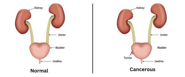
  • Ultrasound Scan: An ultrasound scan uses sound waves to create images of the bladder, kidneys, ureter, and urethra. It helps detect signs of cancer in the bladder and any blockages in the urinary system.
  1. Cystoscopy: This procedure involves inserting a thin tube with a camera (cystoscope) into the bladder to visualize its interior. Biopsies (tissue samples) of the bladder lining may be taken during the cystoscopy to examine for cancer cells.

  2. CT Urogram: A CT urogram combines a CT scan with a contrast dye to examine the kidneys, bladder, and ureters. It provides detailed images and helps determine the stage and location of the cancer.

  3. Blood Tests: Blood tests can assess general health, liver function, kidney function, and blood cell counts, which provide important information about overall health and potential effects of cancer.

  4. MRI Scan: Magnetic resonance imaging (MRI) uses magnetic fields and radio waves to create detailed images of the body. It helps assess the extent of the cancer, including whether it has invaded the deeper layers of the bladder or spread to other parts of the body.

  5. CT Scan: Computed tomography (CT) scan uses x-rays and computer technology to produce cross-sectional images of the body. It can provide information about the location, size, and potential spread of bladder cancer.

  6. PET-CT Scan: A PET-CT scan combines a CT scan with a positron emission tomography (PET) scan. It uses a radioactive tracer to detect areas of increased metabolic activity in the body, helping to evaluate the size, stage, and response to treatment of bladder cancer.

  7. Bone Scan: A bone scan is used to check for any abnormalities or metastasis of bladder cancer in the bones. It may be conducted if there are symptoms suggesting bone involvement.

These tests, conducted by GPs and specialists, play a crucial role in diagnosing bladder cancer, determining its stage, and guiding appropriate treatment decisions.

Treatment

Treatment of Bladder cancer

Treating bladder cancer involves different approaches depending on the stage and risk factors associated with the cancer. Here are the common treatment options:

  • In Non-muscle-Invasive Bladder Cancer (NMIBC) the cancer is limited to the inner lining of the bladder.

Treatment typically starts with a surgical procedure called transurethral resection of a bladder tumor (TURBT). This involves removing cancerous cells while preserving the rest of the bladder.

After TURBT, chemotherapy medication may be instilled directly into the bladder to reduce the risk of cancer recurrence.

In some cases, Bacillus Calmette-Guérin (BCG), a medication that stimulates the immune system, may be injected into the bladder to lower the risk of cancer returning.

  • In Muscle-Invasive Bladder Cancer (MIBC) it invaded the muscle layer of the bladder, and more aggressive treatment is required.

A common approach is surgically removing the entire bladder in a procedure called a cystectomy. After bladder removal, alternative methods for urine collection are necessary.

These can include creating an opening in the abdomen for urine to pass into an external bag or constructing a new bladder using a segment of the bowel.

Cystectomy may be accompanied by other treatments, such as chemotherapy or radiation therapy, to target any remaining cancer cells.

  • Radiation Therapy and Chemotherapy

In cases where bladder removal is not recommended or feasible, a combination of radiation therapy and chemotherapy may be used as primary treatment.

This approach is often employed for individuals who have MIBC or are unfit for surgery. Sometimes chemotherapy is administered prior to surgery or in conjunction with radiation therapy.

Following treatment for any type of bladder cancer, regular follow-up tests will be conducted to monitor for potential recurrence or signs of cancer. These tests are important for ongoing surveillance and ensuring timely intervention if needed.

It is crucial to consult with a healthcare professional who can evaluate the specific characteristics of bladder cancer and guide you towards the most appropriate treatment plan.

Coping with a Bladder Cancer Diagnosis

Coping with Bladder cancer

It can be emotionally challenging, and changes in your body may affect how you feel about yourself. Support is available to help you through this process.

Here are some coping strategies and support options:

Dealing with Your Feelings:

  • Understand that it is normal to have various emotions after a cancer diagnosis. You might feel shocked, upset, numb, frightened, uncertain, confused, angry, resentful, or guilty. Different feelings may come and go, and it’s important to allow yourself time to process and accept your emotions.
  • Gathering information about your type of cancer and treatment can help you feel more informed and empowered. Make a list of questions to ask your doctor and bring someone along to appointments for support and to help remember the information provided.
  • Take things one step at a time and seek help when needed. It’s okay to ask for assistance and support from healthcare professionals, family, and friends.
  • Engage in practical activities like making lists, keeping a calendar of appointments, setting goals, and planning enjoyable activities to help you cope and maintain a sense of control.

Talking to Others:

  • Talking to your friends, relatives, and healthcare providers about your cancer can provide valuable support. However, not everyone may be ready to talk about it, so respect their boundaries and communicate your needs openly.
  • Cancer support groups, helplines, and online forums, such as Cancer Chat, can connect you with people who have had similar experiences and provide additional support.
  • Consider seeing a counselor or therapist who specializes in cancer-related issues to help you navigate the emotional challenges and provide guidance.

Addressing Physical Changes:

  • Bladder cancer and its treatments can lead to physical changes in your body. Discuss any concerns or physical changes with your doctor or specialist nurse, as they can help you manage them effectively.
  • Problems passing urine, changes in appearance (such as scarring or a stoma), fatigue, and the impact on relationships and sex life are common issues. Seek support and information from healthcare professionals who can guide you through these changes and provide resources to cope with them.

Practical Coping:

  • Alongside emotional and physical aspects, it’s important to address practical matters. Financial concerns, work-related issues, childcare, and other practical aspects may need attention. Seek guidance from social workers, financial advisors, or professionals specializing in supporting individuals with cancer.

Remember that you don’t have to face these challenges alone. Reach out to healthcare professionals, support organizations, and your loved ones for assistance. They can provide the support and resources you need to navigate the journey of cancer and its treatment.

Take Care of Yourself!

Also Read

Does Stress make Skin Problem Worse?

Share:

Facebook
WhatsApp
Aviral
Reviewed By : Dr. Aviral Vatsa

Social Media

Most Popular

Get The Latest Updates

Subscribe To Our Weekly Newsletter

No spam, notifications only about new products, updates.

Categories

On Key

Related Posts

Non-Invasive Treatment of Breast Cancer Using Histotripsy

Breast cancer is a global health challenge, with its impact felt across all corners of the world. According to the World Health Organization (WHO), breast cancer is the most frequent cancer among women, impacting 2.1 million women each year. It causes the greatest number of cancer-related deaths among women. In 2020, it is estimated that 685,000 women died from breast cancer worldwide. As the incidence of breast cancer rises, particularly in developing countries where the majority of cases are diagnosed in late stages, the need for accessible and non-invasive treatments is more pressing than ever. Histotripsy promises to be a gamechanger for the non-invasive treatment of breast cancer worldwide. In India, breast cancer has overtaken cervical cancer as the most common cancer among women. The Indian Council of Medical Research (ICMR) has reported a significant rise in cases, with breast cancer accounting for 14% of cancers in Indian women. Urban areas register a higher incidence rate than rural areas, with cities like Delhi, Mumbai, and Kolkata showing increasing numbers. Late diagnosis is a significant issue, with a majority of breast cancer patients presenting at stage 2 or later, which can limit treatment options and reduce survival rates. Globally, the advancements in histotripsy research are paving the way for its potential application in breast cancer treatment. The technology’s precision and the possibility of outpatient procedures could lead to a paradigm shift in cancer care, offering a treatment that is not only effective but also aligns with the needs and preferences of patients. The introduction of histotripsy could be particularly transformative in India, where the healthcare system is burdened by the high volume of patients and often limited resources. The non-invasive nature of histotripsy could provide a less resource-intensive treatment option, reducing the need for surgical facilities and lengthy hospital stays. This could be a game-changer for rural and underserved areas where access to surgical oncologists and specialized care is limited. For countries like India, where cultural factors often influence healthcare decisions, the ability to preserve the breast could lead to increased acceptance and adherence to treatment protocols. The Potential Impact of Histotripsy in Treating Breast Cancer In the context of breast cancer, histotripsy has been shown to effectively reduce tumor burden in both subcutaneous and orthotopic mouse models, indicating its potential for treating primary breast tumors. The immunological responses to histotripsy ablation are mediated by the release of tumor antigens and damage-associated molecular patterns (DAMPs) into the tumor microenvironment. These factors can potentiate the effects of checkpoint inhibition, even sensitizing previously resistant cancers to immunotherapy. Histotripsy has also been shown to promote local and systemic immunological responses, reducing tumor burden in breast cancer. This is achieved by lysing target tumor cells, which release tumor antigens and DAMPs into the tumor microenvironment. The immunomodulatory impact of histotripsy may be key to expanding the impact and promise of cancer immunotherapy. In addition, histotripsy has the potential to be used in combination with other cancer treatments, such as chemotherapy and immunotherapy, to enhance their effectiveness and reduce tumor burden. The immunomodulatory impact of histotripsy may also make it an attractive option for treating metastatic breast cancer, as it can promote the polarization of monocytes and macrophages to pro-anti-tumor phenotypes, reducing the magnitude of tumor-supporting immune cells within the tumor microenvironment. What Is Histotripsy? Histotripsy is a promising non-invasive, non-ionizing, non-thermal ablation modality for the treatment of breast cancer. It initiates local and systemic immunological responses, reduces tumor burden, and promotes an anti-tumor microenvironment. The immunomodulatory impact of histotripsy may be key to expanding the impact and promise of cancer immunotherapy, making it an attractive option for treating primary and metastatic breast cancer. The potential impact of histotripsy in treating breast cancer could be significant due to several key advantages: 1. Non-invasive Treatment Histotripsy’s non-invasive nature stands out as one of its most significant benefits. Traditional cancer surgeries involve incisions, which come with risks such as infection, longer hospital stays, and longer recovery periods. By using focused ultrasound waves, histotripsy can destroy cancerous tissues without making any cuts. This approach can reduce the overall stress on the patient’s body, potentially leading to quicker recovery and less physical trauma. It could be particularly advantageous for patients who are poor candidates for surgery due to other health issues. 2. Precision The accuracy of histotripsy is due to its ability to focus ultrasound waves very precisely on a target area, often with millimeter accuracy. This precision is crucial in the treatment of breast cancer, where the goal is to remove or destroy cancerous cells without damaging the surrounding healthy tissue. Such targeted therapy could lead to better preservation of breast tissue, which is important for the patient’s physical and psychological recovery, potentially improving cosmetic outcomes and reducing the need for reconstructive surgery. 3. Reduced Side Effects Conventional treatments like chemotherapy and radiation can have significant side effects because they are not able to distinguish between healthy and cancerous cells. Histotripsy’s targeted approach could minimize this issue, as the therapy focuses only on the cancerous tissues. This targeted disruption means the body may be less overwhelmed by toxins and the side effects of cell death, possibly resulting in a better quality of life during and after treatment. 4. Repeatable treatment One of the limitations of radiation therapy is that there is a maximum total dose that can be safely administered to a patient. This cap does not exist with histotripsy. Because histotripsy does not use ionizing radiation, treatments can be administered multiple times without the cumulative risks associated with radiation. This repeatability allows clinicians to adjust treatment plans based on how the cancer responds and could be especially beneficial for aggressive or recurring breast cancers. 5. Potential in Drug Delivery Research has suggested that the mechanical disruption of tumor tissues by histotripsy can increase the permeability of those tissues. This change could potentially enhance the delivery and efficacy of chemotherapy drugs by allowing higher concentrations of the drug to penetrate the tumor. Furthermore, the combination of histotripsy and

Non-Invasive Treatment of Liver Tumors: The Revolutionary Role of Histotripsy

Liver tumors, whether benign or malignant, have long been a significant health concern. Traditional treatments, while effective, often involve invasive surgical procedures that come with inherent risks and complications. For many patients, especially those with small, numerous, or strategically located tumors, surgery might not be a viable option. However with Histotripsy, a groundbreaking procedure that promises a non-invasive alternative with remarkable potential, effective non-invasive treatment liver cancer is now possible. Histotripsy is a novel medical procedure that harnesses the power of ultrasound waves to mechanically disintegrate tissue structures. Unlike other treatments that depend on heat or radiation, histotripsy utilizes the sheer force of sound waves. These waves are meticulously focused on the target tissue, leading to its fragmentation without harming the surrounding healthy tissue. The precision and non-invasive nature of histotripsy make it a beacon of hope for liver tumor patients. A Closer Look at Evidence Research has demonstrated the efficacy of histotripsy in creating safe and effective ablations in the in vivo human-scale porcine normal liver. In various studies, target liver volumes ranging from 12–60 ml were completely ablated in durations spanning 20–75 min. The results were consistent and promising: within the ablated region, there was uniform tissue disruption with no viable cells remaining. Impressively, major vessels and bile ducts remained intact. The realm of medical treatments has always been complemented by the power of visual evidence, providing clinicians, researchers, and patients with tangible proof of a procedure’s effectiveness. Magnetic Resonance Imaging (MRI) stands at the forefront of this visual exploration. The high-resolution images produced by MRI scanners offer a detailed look into the internal structures of the liver, both before and after histotripsy treatment. Specifically, axial T2-weighted MR images have been invaluable (Figure 1). These images vividly depict the ablation volume, which is the targeted area that underwent histotripsy. The clarity and precision of these images allow medical professionals to ascertain the exact boundaries of the treated area, ensuring that the tumor cells have been effectively disrupted while leaving the surrounding healthy liver tissues untouched. Complementing the MRI scans are gross morphological examinations. These examinations, often conducted post-procedure, provide a hands-on, macroscopic view of the liver. They reveal a consistent pattern of tissue disruption within the ablation zone, characterized by a lack of viable hepatocytes, which are the primary cells of the liver. This uniformity in tissue disruption is a testament to histotripsy’s precision, ensuring that the treatment is both thorough and targeted. Furthermore, the visual evidence extends beyond just the immediate aftermath of the procedure. MR images from rodent Hepatocellular Carcinoma (HCC) models, a common type of liver cancer, provide insights into the longer-term effects of histotripsy. Initial scans of these models show tumors with a hyperintense signal on T2-weighted MRI, indicating active and aggressive tumor growth. However, post-histotripsy images paint a different picture. The previously hyperintense tumors now display a T2 hypointense signal within the ablation zone, signifying successful disruption of the tumor cells. Even more promising are follow-up scans taken 12 weeks after the procedure. These images often reveal a near-total disappearance of the tumor, replaced by a small fibrous tissue zone. This transformation underscores histotripsy’s potential not just as a treatment method but as a possible path to long-term recovery. The visual evidence supporting histotripsy’s effectiveness is both compelling and comprehensive. From high-resolution MRI scans to hands-on morphological examinations, each piece of evidence builds a case for histotripsy as a revolutionary, non-invasive treatment for liver tumors. What are the benefits of Histotripsy for Liver Tumor Treatment? The Immune Response of Histotripsy Another fascinating aspect of histotripsy is its potential to induce an immune response. FACS analysis of histotripsy-ablated tumors identified significant levels of intratumoral CD8+ T cell infiltration, much higher than untreated control tumors. This suggests that histotripsy might play a role in boosting the body’s natural defenses against cancer cells. Additionally, there was a noticeable reduction in pulmonary metastases in mice treated with histotripsy compared to untreated controls. Given that bones, especially ribs, are highly reflective and absorptive for ultrasound propagation, the feasibility and safety of histotripsy through ribs were meticulously studied. The results were reassuring. Ablation zones created through full ribcage coverage were comparable to those with only overlying soft tissue. The temperature increase to ribs was minimal, ensuring no thermal damage to the ribs or surrounding tissue. However, it’s worth noting that in one paper by Smolock et al., body wall damage was reported. This was likely due to pre-focal cavitation on the ribs. But subsequent studies addressed this by adjusting the focal pressure and duty cycle, effectively eliminating such damage. In some cases, transient thrombosis in portal and hepatic veins was observed within the treatment zone, akin to outcomes from radiofrequency and microwave ablation. The long-term response to liver treatment by histotripsy has also been studied. In normal rodent models, the acellular homogenate generated by histotripsy was absorbed within a month, leaving only a minuscule fibrous region. In tumor treatment studies, tumors were completely absorbed within 7–10 weeks post-histotripsy, with no evidence of residual tumor after three months. In conclusion, Histotripsy is undeniably a game-changer in the realm of liver tumor treatments. Its non-invasive nature, combined with its precision and efficacy, makes it a promising alternative to traditional surgical interventions. As research continues and technology advances, histotripsy could very well become the gold standard for treating not just liver tumors but a plethora of other medical conditions. For patients and medical professionals alike, it heralds a future of safer, more effective, and less invasive treatment options.

Immunological Effects Of Histotripsy for Cancer Therapy

Histotripsy, a groundbreaking non-invasive ultrasonic technique, is rapidly gaining traction in the realm of cancer therapy. By harnessing the power of cavitation, histotripsy meticulously ablates targeted tissues, positioning itself as a potential successor to traditional cancer treatments. This comprehensive article seeks to unravel the intricate immunological responses instigated by histotripsy and its profound implications for cancer therapy. What is the immunological response of Histotripsy? At its core, histotripsy ablation is driven by a phenomenon known as cavitation. This intricate process involves the meticulous generation of microbubbles within the targeted tissues, culminating in their systematic breakdown. As these tissues undergo ablation, cells are fragmented into subcellular components and acellular debris. The immunological aftermath of histotripsy, irrespective of its variant – mHIFU, BH, or CCH, remains consistent, primarily due to the analogous effects they exert on the targeted tissues. While potential nuances between these sub-therapies exist, current research has yet to pinpoint significant disparities in their immunologic outcomes. How does Histotripsy decrease pro-tumor immune cells? The tumor microenvironment is a complex ecosystem comprising various cell types, signaling molecules, and extracellular matrix components. Within this intricate network, certain immune cells, often termed as pro-tumor immune cells, play a pivotal role in promoting tumor growth and metastasis. These cells, which include tumor-associated macrophages (TAMs), myeloid-derived suppressor cells (MDSCs), and regulatory T cells (Tregs), actively suppress anti-tumor immune responses, thereby facilitating tumor progression. a. Tumor-Associated Macrophages (TAMs): TAMs are a subset of macrophages that are recruited to the tumor site and are educated by the tumor microenvironment to adopt a pro-tumor phenotype. These cells can promote tumor growth, angiogenesis, and metastasis while suppressing anti-tumor immune responses. Histotripsy’s ability to target and modulate TAMs is of significant interest. By disrupting the tumor microenvironment, histotripsy can potentially reprogram TAMs from a pro-tumor M2 phenotype to an anti-tumor M1 phenotype, thereby reversing their tumor-promoting effects. b. Myeloid-Derived Suppressor Cells (MDSCs): MDSCs are a heterogeneous group of immature myeloid cells that expand during cancer, inflammation, and infection. In the context of cancer, MDSCs play a crucial role in suppressing T cell responses and promoting tumor growth. Preliminary studies suggest that histotripsy may reduce the number and suppressive function of MDSCs in the tumor microenvironment, thereby enhancing anti-tumor immunity. c. Regulatory T cells (Tregs): Tregs are a subset of CD4+ T cells that play a critical role in maintaining immune homeostasis by suppressing excessive immune responses. However, in the tumor microenvironment, Tregs can inhibit anti-tumor immune responses, thereby facilitating tumor growth. The impact of histotripsy on Tregs is an area of active research. By disrupting the tumor microenvironment and releasing tumor antigens, histotripsy may modulate the function and recruitment of Tregs, potentially enhancing anti-tumor immunity. d. The Interplay with Other Immune Cells: Apart from the aforementioned pro-tumor immune cells, the tumor microenvironment also contains other immune cells like dendritic cells, natural killer cells, and B cells. The interplay between these cells and pro-tumor immune cells is complex and multifaceted. Histotripsy’s ability to modulate this intricate network holds immense therapeutic potential. For instance, by targeting pro-tumor immune cells, histotripsy may enhance the antigen-presenting function of dendritic cells, leading to a more robust activation of T cells and a potent anti-tumor immune response. The tumor microenvironment is a dynamic and complex landscape where various immune cells interact and influence tumor progression. Histotripsy, with its unique mechanism of action, holds the potential to modulate this environment, targeting pro-tumor immune cells, and enhancing anti-tumor immunity. As research in this area continues to evolve, a deeper understanding of histotripsy’s impact on the tumor microenvironment will pave the way for more effective and targeted cancer therapies. What is the role of Damage Associated Molecular Patterns (DAMPs)? Damage Associated Molecular Patterns (DAMPs) are a group of intracellular molecules that have garnered significant attention in the realm of immunology and cellular biology. These molecules, under normal circumstances, reside within the confines of the cell and perform essential functions. However, when cells undergo stress, trauma, or damage, DAMPs are released into the extracellular environment, where they assume a new and critical role. One of the primary functions of DAMPs upon their release is to signal the immune system of potential threats. They act as distress signals, alerting the body to the presence of damaged or dying cells. This is particularly evident in procedures like histotripsy, a non-invasive technique that mechanically disrupts tissue structures. During histotripsy, the cellular damage leads to the liberation of various DAMPs, each with its unique role in the ensuing immune response. For instance, Calreticulin (CRT) is not just a mere bystander in this process. When released, CRT moves to the cell surface, where it aids in antigen presentation. This action effectively marks the damaged cells for elimination, kickstarting an adaptive immune response tailored to address the specific threat.  High Mobility Group  is another pivotal DAMP. As a nuclear protein, its primary role within the cell is to stabilize DNA structures. However, once outside, HMGB1 becomes a potent pro-inflammatory agent. It binds to receptors on immune cells, amplifying the inflammatory response, which is crucial in situations where rapid immune action is needed. Adenosine Triphosphate (ATP), commonly recognized as the primary energy currency of the cell, also plays a role in this immune signaling process. When found outside the cell, ATP acts as a danger signal. Immune cells, recognizing the abnormal presence of extracellular ATP, are prompted to migrate to the site of injury, further intensifying the immune response. Heat Shock Proteins (HSPs) complete the ensemble of key DAMPs released during histotripsy. These proteins, typically produced in response to cellular stress, have a dual role. Inside the cell, they ensure the proper folding of proteins. However, when released, HSPs actively stimulate the immune system, further emphasizing their importance in the body’s defense mechanisms. The intricate connection between DAMPs and the immune response holds profound implications, especially in the field of oncology. Tumors, by their very nature, suppress the immune system, allowing them to grow unchecked. However, the inflammation induced by DAMPs can be harnessed and directed

Histotripsy: A Revolution in Precise Tissue Ablation

Histotripsy has emerged as a beacon of innovation in the ever-evolving landscape of medical technology. It  promises a future where precise tissue ablation can be achieved without the invasiveness of traditional surgical methods. But what makes histotripsy stand out? The answer lies in its unique mechanism of action, which harnesses the power of ultrasound to mechanically disrupt tissue structures. This article delves deep into the mechanism of histotripsy and how it paves the way for precise tissue ablation, especially in the realm of cancer treatment. What is Histotripsy? Histotripsy, a groundbreaking medical technique, is rapidly gaining traction in the healthcare sector due to its potential to revolutionise tissue ablation procedures. The term “histotripsy” is derived from the Greek words “histo,” meaning tissue, and “tripsy,” meaning to break. Histotripsy is a non-invasive approach to tissue disruption, and its unique mechanism of action  sets it apart from any other innovations. . The concept of histotripsy was first introduced at the University of Michigan in 2004. Since its inception, the technique has undergone significant advancements, with researchers continually exploring its potential applications and refining its methodology. The term itself encapsulates the essence of the procedure: a method to break down soft tissue. How does Histotripsy work? The Fundamental Mechanism: Cavitation  Cavitation, a phenomenon central to histotripsy, refers to the formation, growth, and subsequent collapse of gas or vapour-filled bubbles within a liquid medium when subjected to rapid pressure changes. In the context of histotripsy, the human body’s tissue serves as this liquid medium, and high-intensity, short-duration ultrasound pulses induce the rapid pressure changes. When tissues are exposed to these potent ultrasound pulses, the alternating high and low pressures lead to the creation of minuscule bubbles or cavities. These bubbles might initially form around pre-existing gas pockets or microscopic impurities within the tissue. As the ultrasound pulses persist, these bubbles expand due to the negative pressure phases of the ultrasound wave. Bubble Dynamics: The Heart of Tissue Disruption  The true essence of histotripsy is realised during the bubble collapse phase. Following the negative phase, the positive pressure phase of the ultrasound wave causes the expanded bubbles to undergo a swift and violent implosion. This rapid collapse generates potent local shock waves and produces high-velocity liquid jets. These intense mechanical forces, stemming from both the shock waves and the jets, act upon the surrounding tissue. The outcome is a mechanical breakdown of the tissue at a cellular level, resulting in the tissue being fractionated into a liquefied form. This liquid consists of a homogenised blend of cell debris and the extracellular matrix. How does Histotripsy achieve precision in action? In the realm of medical interventions, precision is paramount. The ability to target specific tissues or cells without affecting the surrounding structures can be the difference between successful treatment and unintended complications. Histotripsy, with its groundbreaking approach to tissue ablation, exemplifies this principle of precision in action. Let’s delve deeper into how histotripsy achieves such unparalleled accuracy. Histotripsy employs high-intensity ultrasound pulses to induce cavitation within the targeted tissue. The beauty of this technique lies in the ability to focus these ultrasound beams to a specific point, known as the focal zone. Within this focal zone, the energy of the ultrasound waves is concentrated, ensuring that the cavitation-induced tissue disruption occurs primarily within this localised area. This means that only the tissue within the focal zone is affected, while the surrounding structures remain untouched. One of the standout features that bolster histotripsy’s precision is the integration of real-time imaging. As the ultrasound waves are administered, they not only induce cavitation but also provide a live visual feed of the treatment area. This dual capability allows clinicians to monitor the formation and collapse of bubbles in real-time. Such immediate feedback ensures that the treatment is progressing as intended and allows for on-the-fly adjustments. If, for instance, the bubbles are not forming in the desired location or pattern, the clinician can instantly modify the parameters to achieve the desired effect. The precision of histotripsy can be likened to the accuracy of a surgeon’s scalpel, but without the invasiveness of a blade. The controlled generation and collapse of microbubbles ensure that only the targeted cells or tissues are disrupted. This selectivity is especially crucial when treating tumours or lesions located close to vital organs or critical structures. For example, when targeting a tumour adjacent to a major blood vessel, the precision of histotripsy ensures that the vessel remains unharmed, reducing the risk of bleeding or other complications. In many medical treatments, especially those involving radiation or surgery, there’s always a concern about collateral damage to healthy tissues. Histotripsy’s precision minimises this risk. By confining the tissue disruption to the focal zone, histotripsy ensures that the surrounding healthy tissues are spared. This not only enhances the safety profile of the treatment but also promotes faster healing and recovery. What sets Histotripsy apart from other cancer treatments? Histotripsy’s distinctive non-thermal approach to tissue ablation offers a fresh perspective in the realm of medical interventions. While many therapeutic ultrasound techniques, such as High-Intensity Focused Ultrasound (HIFU), rely on generating heat to achieve therapeutic effects, histotripsy stands apart. Traditional methods work by raising the temperature of the targeted tissue to a point where cellular proteins denature, leading to cell death. Although effective, this thermal approach has inherent risks. Elevated temperatures can inadvertently damage surrounding healthy tissues, especially if the heat spreads beyond the targeted area. Moreover, tissues sensitive to heat, like neural tissues, can be at risk of unintended damage. In contrast, histotripsy operates on a fundamentally different principle. Instead of using heat, it employs mechanical forces to achieve tissue disruption. This is achieved through the controlled generation and violent collapse of microbubbles within the tissue, a process known as cavitation. The implosive collapse of these bubbles generates intense local shock waves and produces high-speed liquid jets. These forces act on the tissue, leading to mechanical breakdown at the cellular level without the need for heat. The non-thermal nature of histotripsy offers