Unlocking the Mystery of Dementia

Dementia-What you need to know

What is Dementia   |   Symptoms   |   Who is Affected   |   How Common   |   Is Memory loss Dementia?   |   Diagnosis   |   Treatment   |   Self Care   |   Risk Factors   |   Medical Advice   |   How Brain is Affected   |   Life Expectancy   |   Important Facts   |

Dementia is a broad term that includes different diseases and conditions affecting how we think, remember, make decisions, and even behave.

When someone has dementia, their mental abilities decline, which can make it hard to do everyday things.

Research shows that around half of people aged 85 and above have some form of dementia. While current medications can slow down the worsening of cognitive problems, we still need better treatments to help people with dementia.

What is Dementia?

Dementia is a term used to describe a decline in mental function, and it’s not a specific disease itself.

When someone has dementia, their mental abilities decrease from a higher level they had before. This decline is significant enough to interfere with their daily life.

People with dementia experience difficulties in various areas, including:

  • Memory
  • Reasoning
  • Language
  • Coordination
  • Mood
  • Behavior

Dementia occurs when certain parts of the brain responsible for learning, memory, decision-making, or language are affected by diseases or infections.

What is Dementia?

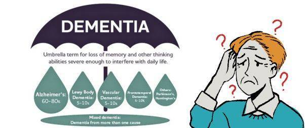
The most common cause of dementia is Alzheimer’s disease.

However, there are other known causes of dementia, including:

  • Vascular dementia
  • Dementia with Lewy bodies
  • Frontotemporal dementia
  • Mixed dementia
  • Dementia due to Parkinson’s disease
  • Dementia-like conditions caused by reversible factors such as medication side effects or thyroid problems.

Signs and Symptoms

In some cases, changes in mood and behavior can occur even before memory problems become apparent. As dementia progresses, symptoms worsen, and most individuals will require assistance with daily activities.

Early signs and symptoms of dementia may include:

  • Forgetting recent events or things
  • Misplacing or losing items
  • Getting lost while walking or driving
  • Feeling confused, even in familiar places
  • Losing track of time
  • Difficulties in problem-solving or decision-making
  • Struggling with conversations or finding the right words
  • Facing challenges in performing familiar tasks
  • Misjudging distances visually

Common changes in mood and behavior can include:

  • Anxiety, sadness, or frustration related to memory loss
  • Personality changes
  • Engaging in inappropriate behaviors
  • Withdrawing from work or social activities
  • Decreased interest in other people’s emotions

Dementia affects each person differently, depending on the underlying causes, existing health conditions, and the individual’s cognitive function prior to the onset of the illness.

Most symptoms tend to worsen over time, although some may disappear or only manifest in later stages of dementia.

As the disease progresses, individuals often require increased assistance with personal care. They may struggle to recognize family members or friends, experience difficulty in mobility, lose control over bodily functions such as bladder and bowel movements, encounter challenges related to eating and drinking, and exhibit behavior changes like aggression that can be distressing for both the person with dementia and those around them.

Who is Affected by Dementia?

Dementia is primarily observed in older individuals, making it a late-life disease.

Approximately 5% to 8% of people aged 65 and above have some form of dementia, and this percentage doubles every five years beyond that age. It is estimated that up to half of individuals aged 85 and older experience dementia.

The prevalence of Alzheimer’s disease and related dementias among different racial groups in the population aged 65 and older is as follows:

  • Blacks: 14%
  • Hispanics: 12%
  • Non-Hispanic whites: 10%
  • American Indian and Alaska Natives: 9%
  • Asian and Pacific Islanders: 8%

These statistics provide insights into the impact of dementia across various racial and ethnic backgrounds within the older population.

How Common is Dementia?

According to the U.S. Centers for Disease Control and Prevention (CDC), approximately 5 million adults aged 65 or older in the United States are currently living with Alzheimer’s disease and related forms of dementia.

Looking ahead, the CDC projects that by the year 2060, the number of people affected by dementia will reach around 14 million, accounting for approximately 3.3% of the population.

Alzheimer’s disease specifically ranks as the sixth leading cause of death in the United States, and it is the fifth leading cause of death among individuals aged 65 and older.

These statistics highlight the significant impact that dementia has on individuals, families, and society as a whole.

Does Memory Loss mean Dementia is Starting?

Does Memory loss is Dementia?

There is a common misconception that memory loss automatically indicates the onset of dementia. However, memory loss can have various causes, and experiencing memory difficulties does not necessarily confirm a dementia diagnosis.

It is important to recognize that certain memory changes are a normal part of aging. As we grow older, some neurons in our brain naturally decline. However, this type of age-related memory loss is not severe enough to significantly impact daily functioning.

Dementia, on the other hand, goes beyond ordinary forgetfulness. It affects a person’s ability to function effectively. Dementia is not simply misplacing keys; it can involve forgetting the purpose of keys altogether. It’s crucial to understand that dementia is not a normal aspect of aging.

It’s essential to differentiate between occasional memory lapses, which can happen to anyone, and persistent memory problems that disrupt daily life. If you or a loved one are concerned about memory loss or cognitive decline, it’s important to consult with a healthcare professional for proper evaluation and diagnosis.

Diagnosing Dementia

Diagnosing dementia can be a complex task as there are various diseases and conditions that can contribute to its development. Moreover, many of the symptoms associated with dementia overlap with those of other illnesses.

To determine whether dementia is the underlying cause of cognitive decline, your healthcare provider will undertake several steps:

  1. Assessing symptom progression: Your healthcare provider will inquire about the pattern and progression of your symptoms to understand how they have evolved over time.
  2. Reviewing medical history: A comprehensive understanding of your medical history is crucial. Your healthcare provider will gather information about your past illnesses, medications, and family history, particularly related to dementia.
  3. Conducting tests: Several tests may be ordered to assist in the diagnostic process, including laboratory tests, imaging tests, and neurocognitive assessments.
    • Laboratory tests: These tests aim to rule out other possible causes of dementia symptoms, such as infections, inflammation, thyroid problems, or vitamin deficiencies.
    • Imaging tests: Brain imaging techniques like computed tomography (CT) or magnetic resonance imaging (MRI) can provide detailed images of the brain, helping to identify abnormalities such as stroke, bleeding, tumors, or fluid accumulation.
    • FDG-PET scan: In certain cases, a specialized brain scan known as FDG-PET may be recommended to evaluate brain function and assess cognitive decline.
  4. Neurocognitive testing: Through written and computerized tests, healthcare providers assess various mental abilities including problem-solving, learning, memory, judgment, planning, reasoning, language, and more.
  5. Psychiatric evaluation: A mental health professional may be involved to evaluate factors like depression, mood changes, or other psychiatric conditions that could contribute to memory loss.

Neurologists and geriatricians often play a crucial role in the diagnostic process, combining their expertise to arrive at an accurate diagnosis.

It’s important to remember that diagnosing dementia requires a comprehensive evaluation and collaboration among healthcare professionals, and the process may take time to ensure an accurate assessment of the condition.

Treatment

When discussing dementia, it’s important to clarify the terms “treatable,” “reversible,” and “curable.” While all or most forms of dementia are considered treatable, meaning that medications and other measures can help manage symptoms, it’s crucial to note that most types of dementia cannot be cured or fully reversed.

Treatment options typically provide modest benefits.

However, there are certain forms of dementia that can be successfully reversed when caused by treatable underlying factors. These dementia-like symptoms may result from:

  • Side effects of medications, illicit drugs, or alcohol
  • Removable tumors
  • Subdural hematoma (blood accumulation beneath the brain’s outer covering caused by head injury)
  • Normal pressure hydrocephalus (excessive cerebrospinal fluid buildup in the brain)
  • Metabolic disorders, such as vitamin B12 deficiency
  • Hypothyroidism (low levels of thyroid hormones)
  • Hypoglycemia (low blood sugar)
  • Depression

Even when dementia is not reversible, there are medications available that can partially address memory loss and behavioral issues associated with certain forms of dementia. These include:

  • Alzheimer’s disease
  • Multi-infarct (vascular) dementia
  • Dementias related to Parkinson’s disease and similar conditions
  • AIDS dementia complex
  • Creutzfeldt-Jakob disease

While treatment options can help manage symptoms and improve quality of life, it is important to acknowledge that they do not provide a complete cure for dementia. Each case is unique, and treatment plans are tailored to individual needs and circumstances.

It is crucial to consult with healthcare professionals to receive an accurate diagnosis, discuss treatment options, and develop a personalized care plan for managing dementia symptoms.

Self Care with Dementia

Self Care of Dementia

If you have been diagnosed with dementia, there are several self-care strategies that can help you manage your symptoms and improve your overall well-being:

  1. Stay physically active: Engage in regular physical activity that suits your abilities and interests. It can be as simple as taking walks, doing light exercises, or participating in activities like yoga or tai chi.
  2. Eat healthily: Maintain a balanced diet rich in fruits, vegetables, whole grains, and lean proteins. Proper nutrition is vital for brain health and overall well-being.
  3. Stop smoking and drinking alcohol: If you smoke or consume alcohol, consider quitting or reducing your intake. Both smoking and excessive alcohol consumption can worsen symptoms and have negative effects on your health.
  4. Regular check-ups: Schedule regular visits with your doctor to monitor your overall health and address any concerns or changes in your condition.
  5. Use memory aids: Help manage memory difficulties by writing down important tasks, appointments, and reminders. Utilize calendars, sticky notes, or smartphone apps to assist you in staying organized.
  6. Engage in enjoyable activities: Continue pursuing hobbies and activities that bring you joy and fulfillment. Participating in activities that stimulate your mind and provide a sense of purpose can be beneficial.
  7. Stay connected: Maintain social connections with friends, family, and your community. Engaging in social interactions and participating in community life can help improve your emotional well-being.
  8. Plan ahead: As dementia progresses, it may become challenging to make important decisions for yourself or manage your finances. Consider the following steps:
    • Identify trustworthy individuals who can support you in decision-making and help you communicate your preferences.
    • Create an advance plan outlining your care and support preferences, ensuring your wishes are known and respected.
    • Carry identification with your address and emergency contact information whenever you leave the house.
  9. Seek support: Reach out to family, friends, and support groups for assistance. Communicate your needs and concerns, and don’t hesitate to ask for help when needed.
  10. Take care of yourself as a caregiver: If you are providing care and support for someone with dementia, remember to prioritize your own well-being. Seek assistance from others, take regular breaks, and practice stress management techniques. Consider joining support groups or seeking professional guidance to navigate the challenges of caregiving.

By implementing these self-care strategies, you can enhance your quality of life and better manage the impact of dementia on your daily life.

Risk Factors

Several factors can increase the risk of developing dementia. It’s essential to be aware of these risk factors to better understand the potential for developing the condition:

  1. Age: Age is the most significant risk factor for dementia. The likelihood of developing dementia increases as you grow older, with the majority of cases affecting individuals over the age of 65.
  2. Family History: Having a family history of dementia, particularly among biological parents or siblings, can increase your own risk of developing the condition. Genetic factors can play a role in certain types of dementia.
  3. Down Syndrome: Individuals with Down syndrome are at a higher risk of developing early-onset Alzheimer’s disease during middle age. The presence of an extra chromosome 21 in individuals with Down syndrome contributes to this increased risk.
  4. Poor Heart Health: Poor cardiovascular health, characterized by conditions like high cholesterol levels, high blood pressure, atherosclerosis, and smoking, can raise the risk of dementia. These conditions, along with diabetes, impact blood vessels, potentially leading to reduced blood flow and an increased likelihood of strokes.
  5. Race and Ethnicity: Studies indicate that certain racial and ethnic groups may have a higher risk of developing dementia. Black individuals have double the risk compared to white individuals, while Hispanic individuals are 1.5 times more likely to develop dementia than their white counterparts.
  6. Brain Injury: Experiencing a severe brain injury can increase the risk of developing dementia. Traumatic brain injuries, such as those resulting from accidents or falls, can have long-term effects on cognitive function.

It’s important to note that having one or more risk factors does not guarantee the development of dementia. Risk factors indicate an increased likelihood but do not provide a definitive prediction.

Many factors contribute to the complex nature of dementia, and additional research is continually being conducted to further understand the risk factors involved.

By being aware of these risk factors, individuals can make informed decisions about their lifestyle, take appropriate preventive measures, and seek early medical intervention if necessary.

When to Seek Medical Advice for Dementia Concerns

When to seek medical advice for Dementia

If you or your loved ones notice any of the following changes, it is advisable to schedule an appointment with your healthcare provider:

  1. Memory: Significant changes in memory, such as forgetting important information, struggling to recall recent events or conversations, or relying heavily on reminders and notes.
  2. Mental Functioning: Noticeable decline in cognitive abilities, including difficulties with problem-solving, decision-making, concentration, or learning new information.
  3. Everyday Tasks: Challenges in performing routine activities that were once familiar and easily accomplished, such as difficulty managing finances, following recipes, or completing household chores.
  4. Behavior: Unusual or uncharacteristic behaviors, such as increased confusion, agitation, aggression, or restlessness that cause concern or disruption in daily life.
  5. Personality: Noticeable changes in personality traits or emotional well-being, including shifts in mood, irritability, apathy, withdrawal from social activities, or loss of interest in hobbies.

It’s important to remember that experiencing one or more of these changes does not necessarily indicate dementia, as various factors can contribute to similar symptoms.

However, discussing your concerns with a healthcare professional will help determine the underlying cause and provide appropriate guidance and support.

How Dementia Impacts the Brain and Body

How Dementia affects Brain and Body

As dementia progresses, the brain deteriorates, leading to memory loss and impaired cognitive function. Basic bodily functions like breathing, digestion, and mobility are affected.

Eventually, individuals with advanced dementia require full-time assistance for essential tasks such as eating, walking, and communicating. They become susceptible to infections and may experience complications like pneumonia.

At this stage, many families opt for hospice care to prioritize comfort and quality of life.

Life Expectancy in Dementia

The life expectancy of a person with dementia can vary significantly depending on several factors.

On average, individuals with dementia live for about 8 to 10 years after the initial symptoms appear. However some individuals may live much longer or shorter lives.

It’s also worth mentioning that dementia is a progressive condition, meaning it worsens over time.

As the disease advances, individuals may experience increased cognitive decline, functional impairment, and other health complications. Quality of life and overall well-being become important considerations in the later stages of dementia.

Ultimately, it’s best to consult with healthcare professionals who can provide personalized information and guidance based on an individual’s specific situation and type of dementia.

Important Facts about Dementia

Important facts about Dementia

  1. Global Impact: Currently, more than 55 million people worldwide have dementia, with over 60% residing in low- and middle-income countries. Each year, nearly 10 million new cases are reported.
  2. Causes and Types: Dementia can be caused by various diseases and brain injuries. The most common form is Alzheimer’s disease, accounting for 60-70% of cases.
  3. Leading Cause: Dementia ranks as the seventh leading cause of death globally and is a major contributor to disability and dependence among older individuals.
  4. Economic Burden: In 2019, dementia cost economies around the world a staggering 1.3 trillion US dollars. Approximately 50% of these costs are attributed to informal caregivers, such as family members and close friends, who provide an average of 5 hours of daily care and supervision.
  5. Gender Disparity: Women are disproportionately affected by dementia, both directly and indirectly. They experience higher levels of disability-adjusted life years and mortality related to dementia. Additionally, women contribute to 70% of the caregiving hours for individuals living with dementia.

Take Care of Yourself!

Also Read

Thunderclap Headaches

Share:

Facebook
WhatsApp
Aviral
Reviewed By : Dr. Aviral Vatsa

Social Media

Most Popular

Get The Latest Updates

Subscribe To Our Weekly Newsletter

No spam, notifications only about new products, updates.

Categories

On Key

Related Posts

Non-Invasive Treatment of Breast Cancer Using Histotripsy

Breast cancer is a global health challenge, with its impact felt across all corners of the world. According to the World Health Organization (WHO), breast cancer is the most frequent cancer among women, impacting 2.1 million women each year. It causes the greatest number of cancer-related deaths among women. In 2020, it is estimated that 685,000 women died from breast cancer worldwide. As the incidence of breast cancer rises, particularly in developing countries where the majority of cases are diagnosed in late stages, the need for accessible and non-invasive treatments is more pressing than ever. Histotripsy promises to be a gamechanger for the non-invasive treatment of breast cancer worldwide. In India, breast cancer has overtaken cervical cancer as the most common cancer among women. The Indian Council of Medical Research (ICMR) has reported a significant rise in cases, with breast cancer accounting for 14% of cancers in Indian women. Urban areas register a higher incidence rate than rural areas, with cities like Delhi, Mumbai, and Kolkata showing increasing numbers. Late diagnosis is a significant issue, with a majority of breast cancer patients presenting at stage 2 or later, which can limit treatment options and reduce survival rates. Globally, the advancements in histotripsy research are paving the way for its potential application in breast cancer treatment. The technology’s precision and the possibility of outpatient procedures could lead to a paradigm shift in cancer care, offering a treatment that is not only effective but also aligns with the needs and preferences of patients. The introduction of histotripsy could be particularly transformative in India, where the healthcare system is burdened by the high volume of patients and often limited resources. The non-invasive nature of histotripsy could provide a less resource-intensive treatment option, reducing the need for surgical facilities and lengthy hospital stays. This could be a game-changer for rural and underserved areas where access to surgical oncologists and specialized care is limited. For countries like India, where cultural factors often influence healthcare decisions, the ability to preserve the breast could lead to increased acceptance and adherence to treatment protocols. The Potential Impact of Histotripsy in Treating Breast Cancer In the context of breast cancer, histotripsy has been shown to effectively reduce tumor burden in both subcutaneous and orthotopic mouse models, indicating its potential for treating primary breast tumors. The immunological responses to histotripsy ablation are mediated by the release of tumor antigens and damage-associated molecular patterns (DAMPs) into the tumor microenvironment. These factors can potentiate the effects of checkpoint inhibition, even sensitizing previously resistant cancers to immunotherapy. Histotripsy has also been shown to promote local and systemic immunological responses, reducing tumor burden in breast cancer. This is achieved by lysing target tumor cells, which release tumor antigens and DAMPs into the tumor microenvironment. The immunomodulatory impact of histotripsy may be key to expanding the impact and promise of cancer immunotherapy. In addition, histotripsy has the potential to be used in combination with other cancer treatments, such as chemotherapy and immunotherapy, to enhance their effectiveness and reduce tumor burden. The immunomodulatory impact of histotripsy may also make it an attractive option for treating metastatic breast cancer, as it can promote the polarization of monocytes and macrophages to pro-anti-tumor phenotypes, reducing the magnitude of tumor-supporting immune cells within the tumor microenvironment. What Is Histotripsy? Histotripsy is a promising non-invasive, non-ionizing, non-thermal ablation modality for the treatment of breast cancer. It initiates local and systemic immunological responses, reduces tumor burden, and promotes an anti-tumor microenvironment. The immunomodulatory impact of histotripsy may be key to expanding the impact and promise of cancer immunotherapy, making it an attractive option for treating primary and metastatic breast cancer. The potential impact of histotripsy in treating breast cancer could be significant due to several key advantages: 1. Non-invasive Treatment Histotripsy’s non-invasive nature stands out as one of its most significant benefits. Traditional cancer surgeries involve incisions, which come with risks such as infection, longer hospital stays, and longer recovery periods. By using focused ultrasound waves, histotripsy can destroy cancerous tissues without making any cuts. This approach can reduce the overall stress on the patient’s body, potentially leading to quicker recovery and less physical trauma. It could be particularly advantageous for patients who are poor candidates for surgery due to other health issues. 2. Precision The accuracy of histotripsy is due to its ability to focus ultrasound waves very precisely on a target area, often with millimeter accuracy. This precision is crucial in the treatment of breast cancer, where the goal is to remove or destroy cancerous cells without damaging the surrounding healthy tissue. Such targeted therapy could lead to better preservation of breast tissue, which is important for the patient’s physical and psychological recovery, potentially improving cosmetic outcomes and reducing the need for reconstructive surgery. 3. Reduced Side Effects Conventional treatments like chemotherapy and radiation can have significant side effects because they are not able to distinguish between healthy and cancerous cells. Histotripsy’s targeted approach could minimize this issue, as the therapy focuses only on the cancerous tissues. This targeted disruption means the body may be less overwhelmed by toxins and the side effects of cell death, possibly resulting in a better quality of life during and after treatment. 4. Repeatable treatment One of the limitations of radiation therapy is that there is a maximum total dose that can be safely administered to a patient. This cap does not exist with histotripsy. Because histotripsy does not use ionizing radiation, treatments can be administered multiple times without the cumulative risks associated with radiation. This repeatability allows clinicians to adjust treatment plans based on how the cancer responds and could be especially beneficial for aggressive or recurring breast cancers. 5. Potential in Drug Delivery Research has suggested that the mechanical disruption of tumor tissues by histotripsy can increase the permeability of those tissues. This change could potentially enhance the delivery and efficacy of chemotherapy drugs by allowing higher concentrations of the drug to penetrate the tumor. Furthermore, the combination of histotripsy and

Non-Invasive Treatment of Liver Tumors: The Revolutionary Role of Histotripsy

Liver tumors, whether benign or malignant, have long been a significant health concern. Traditional treatments, while effective, often involve invasive surgical procedures that come with inherent risks and complications. For many patients, especially those with small, numerous, or strategically located tumors, surgery might not be a viable option. However with Histotripsy, a groundbreaking procedure that promises a non-invasive alternative with remarkable potential, effective non-invasive treatment liver cancer is now possible. Histotripsy is a novel medical procedure that harnesses the power of ultrasound waves to mechanically disintegrate tissue structures. Unlike other treatments that depend on heat or radiation, histotripsy utilizes the sheer force of sound waves. These waves are meticulously focused on the target tissue, leading to its fragmentation without harming the surrounding healthy tissue. The precision and non-invasive nature of histotripsy make it a beacon of hope for liver tumor patients. A Closer Look at Evidence Research has demonstrated the efficacy of histotripsy in creating safe and effective ablations in the in vivo human-scale porcine normal liver. In various studies, target liver volumes ranging from 12–60 ml were completely ablated in durations spanning 20–75 min. The results were consistent and promising: within the ablated region, there was uniform tissue disruption with no viable cells remaining. Impressively, major vessels and bile ducts remained intact. The realm of medical treatments has always been complemented by the power of visual evidence, providing clinicians, researchers, and patients with tangible proof of a procedure’s effectiveness. Magnetic Resonance Imaging (MRI) stands at the forefront of this visual exploration. The high-resolution images produced by MRI scanners offer a detailed look into the internal structures of the liver, both before and after histotripsy treatment. Specifically, axial T2-weighted MR images have been invaluable (Figure 1). These images vividly depict the ablation volume, which is the targeted area that underwent histotripsy. The clarity and precision of these images allow medical professionals to ascertain the exact boundaries of the treated area, ensuring that the tumor cells have been effectively disrupted while leaving the surrounding healthy liver tissues untouched. Complementing the MRI scans are gross morphological examinations. These examinations, often conducted post-procedure, provide a hands-on, macroscopic view of the liver. They reveal a consistent pattern of tissue disruption within the ablation zone, characterized by a lack of viable hepatocytes, which are the primary cells of the liver. This uniformity in tissue disruption is a testament to histotripsy’s precision, ensuring that the treatment is both thorough and targeted. Furthermore, the visual evidence extends beyond just the immediate aftermath of the procedure. MR images from rodent Hepatocellular Carcinoma (HCC) models, a common type of liver cancer, provide insights into the longer-term effects of histotripsy. Initial scans of these models show tumors with a hyperintense signal on T2-weighted MRI, indicating active and aggressive tumor growth. However, post-histotripsy images paint a different picture. The previously hyperintense tumors now display a T2 hypointense signal within the ablation zone, signifying successful disruption of the tumor cells. Even more promising are follow-up scans taken 12 weeks after the procedure. These images often reveal a near-total disappearance of the tumor, replaced by a small fibrous tissue zone. This transformation underscores histotripsy’s potential not just as a treatment method but as a possible path to long-term recovery. The visual evidence supporting histotripsy’s effectiveness is both compelling and comprehensive. From high-resolution MRI scans to hands-on morphological examinations, each piece of evidence builds a case for histotripsy as a revolutionary, non-invasive treatment for liver tumors. What are the benefits of Histotripsy for Liver Tumor Treatment? The Immune Response of Histotripsy Another fascinating aspect of histotripsy is its potential to induce an immune response. FACS analysis of histotripsy-ablated tumors identified significant levels of intratumoral CD8+ T cell infiltration, much higher than untreated control tumors. This suggests that histotripsy might play a role in boosting the body’s natural defenses against cancer cells. Additionally, there was a noticeable reduction in pulmonary metastases in mice treated with histotripsy compared to untreated controls. Given that bones, especially ribs, are highly reflective and absorptive for ultrasound propagation, the feasibility and safety of histotripsy through ribs were meticulously studied. The results were reassuring. Ablation zones created through full ribcage coverage were comparable to those with only overlying soft tissue. The temperature increase to ribs was minimal, ensuring no thermal damage to the ribs or surrounding tissue. However, it’s worth noting that in one paper by Smolock et al., body wall damage was reported. This was likely due to pre-focal cavitation on the ribs. But subsequent studies addressed this by adjusting the focal pressure and duty cycle, effectively eliminating such damage. In some cases, transient thrombosis in portal and hepatic veins was observed within the treatment zone, akin to outcomes from radiofrequency and microwave ablation. The long-term response to liver treatment by histotripsy has also been studied. In normal rodent models, the acellular homogenate generated by histotripsy was absorbed within a month, leaving only a minuscule fibrous region. In tumor treatment studies, tumors were completely absorbed within 7–10 weeks post-histotripsy, with no evidence of residual tumor after three months. In conclusion, Histotripsy is undeniably a game-changer in the realm of liver tumor treatments. Its non-invasive nature, combined with its precision and efficacy, makes it a promising alternative to traditional surgical interventions. As research continues and technology advances, histotripsy could very well become the gold standard for treating not just liver tumors but a plethora of other medical conditions. For patients and medical professionals alike, it heralds a future of safer, more effective, and less invasive treatment options.

Immunological Effects Of Histotripsy for Cancer Therapy

Histotripsy, a groundbreaking non-invasive ultrasonic technique, is rapidly gaining traction in the realm of cancer therapy. By harnessing the power of cavitation, histotripsy meticulously ablates targeted tissues, positioning itself as a potential successor to traditional cancer treatments. This comprehensive article seeks to unravel the intricate immunological responses instigated by histotripsy and its profound implications for cancer therapy. What is the immunological response of Histotripsy? At its core, histotripsy ablation is driven by a phenomenon known as cavitation. This intricate process involves the meticulous generation of microbubbles within the targeted tissues, culminating in their systematic breakdown. As these tissues undergo ablation, cells are fragmented into subcellular components and acellular debris. The immunological aftermath of histotripsy, irrespective of its variant – mHIFU, BH, or CCH, remains consistent, primarily due to the analogous effects they exert on the targeted tissues. While potential nuances between these sub-therapies exist, current research has yet to pinpoint significant disparities in their immunologic outcomes. How does Histotripsy decrease pro-tumor immune cells? The tumor microenvironment is a complex ecosystem comprising various cell types, signaling molecules, and extracellular matrix components. Within this intricate network, certain immune cells, often termed as pro-tumor immune cells, play a pivotal role in promoting tumor growth and metastasis. These cells, which include tumor-associated macrophages (TAMs), myeloid-derived suppressor cells (MDSCs), and regulatory T cells (Tregs), actively suppress anti-tumor immune responses, thereby facilitating tumor progression. a. Tumor-Associated Macrophages (TAMs): TAMs are a subset of macrophages that are recruited to the tumor site and are educated by the tumor microenvironment to adopt a pro-tumor phenotype. These cells can promote tumor growth, angiogenesis, and metastasis while suppressing anti-tumor immune responses. Histotripsy’s ability to target and modulate TAMs is of significant interest. By disrupting the tumor microenvironment, histotripsy can potentially reprogram TAMs from a pro-tumor M2 phenotype to an anti-tumor M1 phenotype, thereby reversing their tumor-promoting effects. b. Myeloid-Derived Suppressor Cells (MDSCs): MDSCs are a heterogeneous group of immature myeloid cells that expand during cancer, inflammation, and infection. In the context of cancer, MDSCs play a crucial role in suppressing T cell responses and promoting tumor growth. Preliminary studies suggest that histotripsy may reduce the number and suppressive function of MDSCs in the tumor microenvironment, thereby enhancing anti-tumor immunity. c. Regulatory T cells (Tregs): Tregs are a subset of CD4+ T cells that play a critical role in maintaining immune homeostasis by suppressing excessive immune responses. However, in the tumor microenvironment, Tregs can inhibit anti-tumor immune responses, thereby facilitating tumor growth. The impact of histotripsy on Tregs is an area of active research. By disrupting the tumor microenvironment and releasing tumor antigens, histotripsy may modulate the function and recruitment of Tregs, potentially enhancing anti-tumor immunity. d. The Interplay with Other Immune Cells: Apart from the aforementioned pro-tumor immune cells, the tumor microenvironment also contains other immune cells like dendritic cells, natural killer cells, and B cells. The interplay between these cells and pro-tumor immune cells is complex and multifaceted. Histotripsy’s ability to modulate this intricate network holds immense therapeutic potential. For instance, by targeting pro-tumor immune cells, histotripsy may enhance the antigen-presenting function of dendritic cells, leading to a more robust activation of T cells and a potent anti-tumor immune response. The tumor microenvironment is a dynamic and complex landscape where various immune cells interact and influence tumor progression. Histotripsy, with its unique mechanism of action, holds the potential to modulate this environment, targeting pro-tumor immune cells, and enhancing anti-tumor immunity. As research in this area continues to evolve, a deeper understanding of histotripsy’s impact on the tumor microenvironment will pave the way for more effective and targeted cancer therapies. What is the role of Damage Associated Molecular Patterns (DAMPs)? Damage Associated Molecular Patterns (DAMPs) are a group of intracellular molecules that have garnered significant attention in the realm of immunology and cellular biology. These molecules, under normal circumstances, reside within the confines of the cell and perform essential functions. However, when cells undergo stress, trauma, or damage, DAMPs are released into the extracellular environment, where they assume a new and critical role. One of the primary functions of DAMPs upon their release is to signal the immune system of potential threats. They act as distress signals, alerting the body to the presence of damaged or dying cells. This is particularly evident in procedures like histotripsy, a non-invasive technique that mechanically disrupts tissue structures. During histotripsy, the cellular damage leads to the liberation of various DAMPs, each with its unique role in the ensuing immune response. For instance, Calreticulin (CRT) is not just a mere bystander in this process. When released, CRT moves to the cell surface, where it aids in antigen presentation. This action effectively marks the damaged cells for elimination, kickstarting an adaptive immune response tailored to address the specific threat.  High Mobility Group  is another pivotal DAMP. As a nuclear protein, its primary role within the cell is to stabilize DNA structures. However, once outside, HMGB1 becomes a potent pro-inflammatory agent. It binds to receptors on immune cells, amplifying the inflammatory response, which is crucial in situations where rapid immune action is needed. Adenosine Triphosphate (ATP), commonly recognized as the primary energy currency of the cell, also plays a role in this immune signaling process. When found outside the cell, ATP acts as a danger signal. Immune cells, recognizing the abnormal presence of extracellular ATP, are prompted to migrate to the site of injury, further intensifying the immune response. Heat Shock Proteins (HSPs) complete the ensemble of key DAMPs released during histotripsy. These proteins, typically produced in response to cellular stress, have a dual role. Inside the cell, they ensure the proper folding of proteins. However, when released, HSPs actively stimulate the immune system, further emphasizing their importance in the body’s defense mechanisms. The intricate connection between DAMPs and the immune response holds profound implications, especially in the field of oncology. Tumors, by their very nature, suppress the immune system, allowing them to grow unchecked. However, the inflammation induced by DAMPs can be harnessed and directed

Histotripsy: A Revolution in Precise Tissue Ablation

Histotripsy has emerged as a beacon of innovation in the ever-evolving landscape of medical technology. It  promises a future where precise tissue ablation can be achieved without the invasiveness of traditional surgical methods. But what makes histotripsy stand out? The answer lies in its unique mechanism of action, which harnesses the power of ultrasound to mechanically disrupt tissue structures. This article delves deep into the mechanism of histotripsy and how it paves the way for precise tissue ablation, especially in the realm of cancer treatment. What is Histotripsy? Histotripsy, a groundbreaking medical technique, is rapidly gaining traction in the healthcare sector due to its potential to revolutionise tissue ablation procedures. The term “histotripsy” is derived from the Greek words “histo,” meaning tissue, and “tripsy,” meaning to break. Histotripsy is a non-invasive approach to tissue disruption, and its unique mechanism of action  sets it apart from any other innovations. . The concept of histotripsy was first introduced at the University of Michigan in 2004. Since its inception, the technique has undergone significant advancements, with researchers continually exploring its potential applications and refining its methodology. The term itself encapsulates the essence of the procedure: a method to break down soft tissue. How does Histotripsy work? The Fundamental Mechanism: Cavitation  Cavitation, a phenomenon central to histotripsy, refers to the formation, growth, and subsequent collapse of gas or vapour-filled bubbles within a liquid medium when subjected to rapid pressure changes. In the context of histotripsy, the human body’s tissue serves as this liquid medium, and high-intensity, short-duration ultrasound pulses induce the rapid pressure changes. When tissues are exposed to these potent ultrasound pulses, the alternating high and low pressures lead to the creation of minuscule bubbles or cavities. These bubbles might initially form around pre-existing gas pockets or microscopic impurities within the tissue. As the ultrasound pulses persist, these bubbles expand due to the negative pressure phases of the ultrasound wave. Bubble Dynamics: The Heart of Tissue Disruption  The true essence of histotripsy is realised during the bubble collapse phase. Following the negative phase, the positive pressure phase of the ultrasound wave causes the expanded bubbles to undergo a swift and violent implosion. This rapid collapse generates potent local shock waves and produces high-velocity liquid jets. These intense mechanical forces, stemming from both the shock waves and the jets, act upon the surrounding tissue. The outcome is a mechanical breakdown of the tissue at a cellular level, resulting in the tissue being fractionated into a liquefied form. This liquid consists of a homogenised blend of cell debris and the extracellular matrix. How does Histotripsy achieve precision in action? In the realm of medical interventions, precision is paramount. The ability to target specific tissues or cells without affecting the surrounding structures can be the difference between successful treatment and unintended complications. Histotripsy, with its groundbreaking approach to tissue ablation, exemplifies this principle of precision in action. Let’s delve deeper into how histotripsy achieves such unparalleled accuracy. Histotripsy employs high-intensity ultrasound pulses to induce cavitation within the targeted tissue. The beauty of this technique lies in the ability to focus these ultrasound beams to a specific point, known as the focal zone. Within this focal zone, the energy of the ultrasound waves is concentrated, ensuring that the cavitation-induced tissue disruption occurs primarily within this localised area. This means that only the tissue within the focal zone is affected, while the surrounding structures remain untouched. One of the standout features that bolster histotripsy’s precision is the integration of real-time imaging. As the ultrasound waves are administered, they not only induce cavitation but also provide a live visual feed of the treatment area. This dual capability allows clinicians to monitor the formation and collapse of bubbles in real-time. Such immediate feedback ensures that the treatment is progressing as intended and allows for on-the-fly adjustments. If, for instance, the bubbles are not forming in the desired location or pattern, the clinician can instantly modify the parameters to achieve the desired effect. The precision of histotripsy can be likened to the accuracy of a surgeon’s scalpel, but without the invasiveness of a blade. The controlled generation and collapse of microbubbles ensure that only the targeted cells or tissues are disrupted. This selectivity is especially crucial when treating tumours or lesions located close to vital organs or critical structures. For example, when targeting a tumour adjacent to a major blood vessel, the precision of histotripsy ensures that the vessel remains unharmed, reducing the risk of bleeding or other complications. In many medical treatments, especially those involving radiation or surgery, there’s always a concern about collateral damage to healthy tissues. Histotripsy’s precision minimises this risk. By confining the tissue disruption to the focal zone, histotripsy ensures that the surrounding healthy tissues are spared. This not only enhances the safety profile of the treatment but also promotes faster healing and recovery. What sets Histotripsy apart from other cancer treatments? Histotripsy’s distinctive non-thermal approach to tissue ablation offers a fresh perspective in the realm of medical interventions. While many therapeutic ultrasound techniques, such as High-Intensity Focused Ultrasound (HIFU), rely on generating heat to achieve therapeutic effects, histotripsy stands apart. Traditional methods work by raising the temperature of the targeted tissue to a point where cellular proteins denature, leading to cell death. Although effective, this thermal approach has inherent risks. Elevated temperatures can inadvertently damage surrounding healthy tissues, especially if the heat spreads beyond the targeted area. Moreover, tissues sensitive to heat, like neural tissues, can be at risk of unintended damage. In contrast, histotripsy operates on a fundamentally different principle. Instead of using heat, it employs mechanical forces to achieve tissue disruption. This is achieved through the controlled generation and violent collapse of microbubbles within the tissue, a process known as cavitation. The implosive collapse of these bubbles generates intense local shock waves and produces high-speed liquid jets. These forces act on the tissue, leading to mechanical breakdown at the cellular level without the need for heat. The non-thermal nature of histotripsy offers