Shedding Light on the Truth About Cancer: Debunking Common Misconceptions

Highlights

In an age where information overload is the norm, it can be challenging to separate fact from fiction, especially when it comes to health-related topics like Cancer.

With the internet serving as a breeding ground for misinformation, scare stories about cancer can spread rapidly through well-intentioned shares on social media and email chains.

Unfortunately, some of these stories may be fueled by companies aiming to promote their “safer” or “chemical-free” products, leading to widespread confusion and anxiety.

With millions of people affected by cancer worldwide, it is crucial to debunk these myths and provide accurate information to raise awareness and promote informed decision-making.

In this article, we debunk some of the most prevalent cancer myths so that you can focus on what truly matters.

Myth: Cancer is Contagious

Cancer is contagious-Debunking the Myth

Fact: Cancer is not contagious and cannot be transmitted from person to person through touch, bodily fluids, or any other means.

It is caused by genetic mutations within cells, which can be influenced by a combination of genetic, environmental, and lifestyle factors.

Myth: Cancer is Always a Death Sentence

Cancer is not always a death sentence

Fact: Cancer research, diagnosis, and treatment advances have significantly improved survival rates over the past few decades. Early detection and appropriate treatment can lead to favorable outcomes for many cancer patients.

It is important to remember that cancer is not a single disease but a collection of over 100 different types, each with its own characteristics and prognosis.

Myth: Cancer is Always Caused by an Unhealthy Lifestyle

Cancer is because of unhealthy lifestyle-Debunking the Myth

Fact: While lifestyle choices can indeed contribute to cancer risk, they are not the sole cause. Many factors, including genetics, environmental exposures, and random mutations during cell division, can lead to cancer.

Although adopting a healthy lifestyle is important for cancer prevention, it does not guarantee that an individual will never develop the disease.

Myth: Antiperspirants and Underwired Bras Cause Breast Cancer

Antiperspirants and underwired bras cause breast cancer

Fact: For nearly two decades, claims suggesting that antiperspirants and underwired bras cause breast cancer have persisted. However, you can breathe a sigh of relief knowing that no scientific evidence supports these claims. The theories behind these claims include:

  1. Antiperspirants prevent the body from eliminating “toxins,” leading to cancer.
  2. Chemicals from antiperspirants enter the lymph nodes through sweat glands.
  3. Bras, especially those with underwires, block lymph channels, impairing the body’s ability to fight off cancer and infection.

Despite the prevalence of these theories, there is no scientific evidence, anatomical basis, or medical foundation to support them.

It is worth noting that over half of the breast cancers are found in the quarter of the breast closest to the armpit, but this is simply because that area contains most of the breast tissue.

Myth: Bubble Bath Causes Cancer

Bubble bath causes cancer

Sodium lauryl sulfate (SLS) is a widely used ingredient in bubble baths, shower gels, and shampoos, and has been the subject of cancer-related concerns in recent years. However, it’s essential to separate fact from fiction when it comes to the safety of this common ingredient.

Fact: There is no scientific evidence to suggest that SLS has any cancer-causing effects. The concerns surrounding SLS can be traced back to a single website and its purported “author” in America, who claimed that his name was being misused on the internet by companies promoting “natural” bath products.

This revelation confirms that the claims about SLS causing cancer are unfounded and driven by marketing tactics rather than genuine research.

While SLS is a highly effective detergent and is used in higher concentrations for industrial cleaning products, it is not a carcinogen.

However, it can cause skin and eye irritation, especially at high concentrations. If you have dry skin or hair, you may want to avoid products containing SLS, but there is no need to worry about cancer risk.

Myth: Microwaving Food in Plastic Containers is dangerous and Causes Cancer

Plastic containers in microwave cause cancer

Microwaving food in plastic containers has long been a subject of concern for some people, who believe that it could pose a danger to our health.

However, careful investigation and research have provided evidence to counter these claims.

The thorough investigation by the Food and Drug Administration (FDA) regarding the safety of microwaving food in plastic containers says “Any trace amounts of plastic that might dissolve while heating food are well within safe limits“.

Moreover, the FDA also examined claims that plastic containers and clingfilm food wrapping contain dioxins, which have been linked to cancer. The results showed that there are no dioxins present in any plastic container or clingfilm food wrapping.

The convenience of using microwave ovens and plastic containers can be enjoyed without fear, as research has debunked the concerns surrounding their use.

Myth: Mobile Phone use causes Cancer

Mobile Phone causes Cancer-MythConcerns about the potential link between mobile phone use and cancer have circulated for years, with many wondering if their devices could put their health at risk.

Fact: The majority of studies on mobile phone use, including a massive study involving over 400,000 participants, with 50,000 using mobile phones regularly for more than ten years, have found no link between mobile phone use and cancer development.

However, one study has suggested that long-term mobile phone use (more than ten years) might increase the risk of a non-cancerous tumor of the hearing nerve (acoustic neuroma) and a rare but aggressive brain cancer called Glioma.

Despite these findings, the evidence is not strong enough to warrant discarding mobile phones altogether.

While the evidence connecting mobile phone use to cancer is inconclusive and mostly reassuring, you may still want to consider taking some precautions. Limiting the time spent talking on your mobile or using a headset that keeps the antenna away from your brain are potential measures to reduce any potential risk.

Myth: Talcum Powder causes Ovarian Cancer

Talcum Powder causes cancer-Myth

In 2016, talcum powder made global headlines when Johnson & Johnson was ordered to pay over £40 million in compensation to an American woman who developed ovarian cancer. This incident has raised concerns about the potential link between talcum powder and cancer.

Fact: A study involving 4,000 women (half of whom had ovarian cancer) concluded that using talc was associated with a 33% higher risk of developing the disease, with a trend for increasing risk depending on the duration and frequency of use.

However, this study had several flaws, and the number of women affected was minimal – even among those at the highest risk, only 41 cases of ovarian cancer were reported over 24 years.

Despite these findings, it is essential not to panic if you have used talcum powder in the past. The evidence is inconclusive, and the overall risk remains relatively low.

There are no known risks associated with avoiding talcum powder, and adopting a “better safe than sorry” approach may provide peace of mind.

Myth: Pesticides in Food Cause Cancer

Do Pesticides in Food cause Cancer?

Fears surrounding the presence of pesticides in food and their potential link to cancer have persisted for years, often stemming from the use of potent chemicals on crops in the past.

Fact: While concerns about pesticides and cancer risk have historical roots, modern pesticides are carefully regulated and monitored. National guidelines suggest that any potential risk, if present, is vastly outweighed by the health benefits of consuming fresh fruits and vegetables.

Nevertheless, it is always prudent to take precautionary measures. Washing your fruits and vegetables thoroughly before consumption can help minimize any potential risks associated with pesticide residues.

Myth: Artificial Sweeteners cause Cancer

Do Artificial Sweeteners cause Cancer

The safety of artificial sweeteners, such as aspartame, has been a subject of debate and concern for many, with some suggesting a potential link to cancer.

Fact: The best available evidence, including numerous studies and reviews, demonstrates that artificial sweeteners, including aspartame, do not increase the risk of cancer.

The European Food Safety Authority (EFSA) has also reviewed the evidence, concluding that artificial sweeteners are not harmful within daily limits, which are more than most people would consume in one day.

Artificial sweeteners are chemical substances that taste sweet and are used as sugar substitutes in various products, including soft drinks, desserts, chewing gum, and toothpaste. They provide a sweet taste with fewer or no calories and are often found in “diet” or low/zero sugar products.

No link has been found between artificial sweeteners in diet or zero-sugar fizzy drinks and cancer. Low-calorie drinks containing artificial sweeteners can be part of a healthy, balanced diet.

It is essential to focus on proven causes of cancer and adopt a healthy lifestyle to reduce your risk.

Myth: Eating burnt food causes cancer

Do burnt food cause cancer?

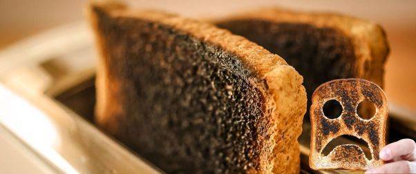
A common concern is whether eating burnt foods or foods high in acrylamide, such as toast or charred root vegetables, can increase the risk of cancer.

Fact: Acrylamide is a chemical found in starchy foods like bread and potatoes when cooked at high temperatures for extended periods.

However, the best available evidence does not show a link between consuming acrylamide from burnt toast, burnt chips, or crispy potatoes and an increased risk of cancer. High-quality studies have not demonstrated that acrylamide from food causes cancer in humans.

Instead, it is essential to focus on maintaining a healthy, balanced diet, which includes more fruits and vegetables, high-fiber foods, and reduced consumption of processed and red meats, sugars, fats, and salt.

Reducing processed meat consumption is a good idea for reducing cancer risk.

Although concerns exist about acrylamide and burnt foods, the best evidence available does not support a connection between these foods and increased cancer risk.

Myth: Milk and Dairy Products cause Cancer

Do Milk & Dairy Products cause Cancer?

Many people wonder if consuming milk and dairy products can cause cancer

Fact: There is insufficient evidence to prove that milk and dairy products cause cancer.

Casein, the primary protein in milk, has not been shown to cause cancer in humans.

Dairy products contain essential proteins, vitamins, and calcium that contribute to overall health, and high calcium content could play a role in reducing bowel cancer risk.

Dairy products do contain some hormones, but the amounts are very small compared to what the body naturally produces, and there is no strong evidence to show that these hormones cause cancer.

The NHS Eatwell Guide recommends including some dairy or dairy alternatives as part of a healthy, balanced diet.

Myth: An Injury or Blow to the Breast causes Cancer

Do Injury & Blow cause Breast Cancer?

Fact: Injuries, trauma, or blows to the breast do not cause cancer. There is no good explanation for how an injury could lead to cancer in breast tissue. However, injuries may sometimes lead to the discovery of cancer that was already present near the injured area.

If someone experiences an injury or blow to the breast, they may check the area themselves or have it examined by a healthcare professional.

If there is already cancer present, these checks could detect it, but this does not mean the injury caused the cancer. While they may sometimes lead to the discovery of pre-existing cancer or the formation of non-cancerous lumps.

Myth: Cosmetics Cause Cancer

Do Cosmetics cause Cancer?

There are concerns about whether chemicals in cosmetics can cause cancer.

Fact: There is no evidence that antiperspirants, or body sprays containing aluminum cause cancer. Personal use of hair dye is not linked to increased cancer risk, although daily exposure in hairdressers and barbers might raise bladder cancer risk, requiring further research.

Parabens used as preservatives in cosmetics, don’t cause cancer, and their potential estrogenic effect in rats hasn’t been proven in humans.

Myth: Medical Scans and Air Travel cause Cancer

Do Medical Scans Cause Cancer?

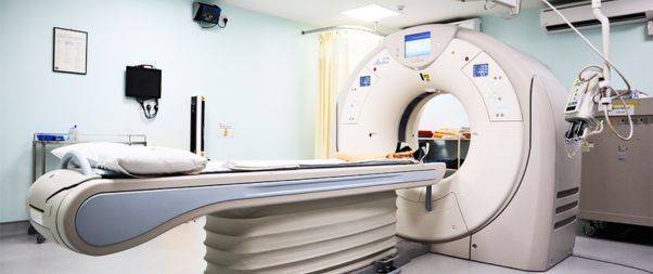
High-energy ionizing radiation can cause cancer by damaging cells and DNA. Some medical scans, like X-rays, expose us to small amounts of ionizing radiation, but the health risks are low and typically outweighed by the benefits of accurate diagnosis.

Radiation exposure during air travel or airport body scanners is unlikely to increase cancer risk.

Cosmic radiation is slightly higher at higher altitudes, but the additional exposure for passengers is minimal and unlikely to affect cancer risk, even for frequent flyers.

Airport body scanners use low levels of ionizing radiation or non-ionizing radiation, which have not been shown to increase cancer risk.

Myth: Stress cause cancer

Do Stress Causes Cancer?

Stress does not directly increase cancer risk. High-quality, long-term studies have found no evidence linking stress to a higher risk of cancer.

However, how people cope with or manage stress can affect their health and potentially impact cancer risk.

Stress may lead to unhealthy behaviors such as smoking, poor diet, excessive alcohol consumption, or reduced physical activity, which can indirectly increase cancer risk.

If you are struggling with prolonged stress, it’s important to talk to your doctor about ways to manage it. Remember, there are many proven ways to reduce cancer risk and maintain a healthy lifestyle.

In a Nutshell

Dispelling cancer myths is crucial for providing accurate information to the public and empowering individuals to make informed decisions about their health.

By debunking these misconceptions, we can promote greater understanding and encourage proactive steps toward cancer prevention and early detection.

Always consult with a healthcare professional for personalized advice and evidence-based information on cancer and its treatments.

Take Care of Yourself!

Share:

Facebook
WhatsApp
Aviral
Reviewed By : Dr. Aviral Vatsa

Social Media

Most Popular

Get The Latest Updates

Subscribe To Our Weekly Newsletter

No spam, notifications only about new products, updates.

Categories

On Key

Related Posts

Non-Invasive Treatment of Breast Cancer Using Histotripsy

Breast cancer is a global health challenge, with its impact felt across all corners of the world. According to the World Health Organization (WHO), breast cancer is the most frequent cancer among women, impacting 2.1 million women each year. It causes the greatest number of cancer-related deaths among women. In 2020, it is estimated that 685,000 women died from breast cancer worldwide. As the incidence of breast cancer rises, particularly in developing countries where the majority of cases are diagnosed in late stages, the need for accessible and non-invasive treatments is more pressing than ever. Histotripsy promises to be a gamechanger for the non-invasive treatment of breast cancer worldwide. In India, breast cancer has overtaken cervical cancer as the most common cancer among women. The Indian Council of Medical Research (ICMR) has reported a significant rise in cases, with breast cancer accounting for 14% of cancers in Indian women. Urban areas register a higher incidence rate than rural areas, with cities like Delhi, Mumbai, and Kolkata showing increasing numbers. Late diagnosis is a significant issue, with a majority of breast cancer patients presenting at stage 2 or later, which can limit treatment options and reduce survival rates. Globally, the advancements in histotripsy research are paving the way for its potential application in breast cancer treatment. The technology’s precision and the possibility of outpatient procedures could lead to a paradigm shift in cancer care, offering a treatment that is not only effective but also aligns with the needs and preferences of patients. The introduction of histotripsy could be particularly transformative in India, where the healthcare system is burdened by the high volume of patients and often limited resources. The non-invasive nature of histotripsy could provide a less resource-intensive treatment option, reducing the need for surgical facilities and lengthy hospital stays. This could be a game-changer for rural and underserved areas where access to surgical oncologists and specialized care is limited. For countries like India, where cultural factors often influence healthcare decisions, the ability to preserve the breast could lead to increased acceptance and adherence to treatment protocols. The Potential Impact of Histotripsy in Treating Breast Cancer In the context of breast cancer, histotripsy has been shown to effectively reduce tumor burden in both subcutaneous and orthotopic mouse models, indicating its potential for treating primary breast tumors. The immunological responses to histotripsy ablation are mediated by the release of tumor antigens and damage-associated molecular patterns (DAMPs) into the tumor microenvironment. These factors can potentiate the effects of checkpoint inhibition, even sensitizing previously resistant cancers to immunotherapy. Histotripsy has also been shown to promote local and systemic immunological responses, reducing tumor burden in breast cancer. This is achieved by lysing target tumor cells, which release tumor antigens and DAMPs into the tumor microenvironment. The immunomodulatory impact of histotripsy may be key to expanding the impact and promise of cancer immunotherapy. In addition, histotripsy has the potential to be used in combination with other cancer treatments, such as chemotherapy and immunotherapy, to enhance their effectiveness and reduce tumor burden. The immunomodulatory impact of histotripsy may also make it an attractive option for treating metastatic breast cancer, as it can promote the polarization of monocytes and macrophages to pro-anti-tumor phenotypes, reducing the magnitude of tumor-supporting immune cells within the tumor microenvironment. What Is Histotripsy? Histotripsy is a promising non-invasive, non-ionizing, non-thermal ablation modality for the treatment of breast cancer. It initiates local and systemic immunological responses, reduces tumor burden, and promotes an anti-tumor microenvironment. The immunomodulatory impact of histotripsy may be key to expanding the impact and promise of cancer immunotherapy, making it an attractive option for treating primary and metastatic breast cancer. The potential impact of histotripsy in treating breast cancer could be significant due to several key advantages: 1. Non-invasive Treatment Histotripsy’s non-invasive nature stands out as one of its most significant benefits. Traditional cancer surgeries involve incisions, which come with risks such as infection, longer hospital stays, and longer recovery periods. By using focused ultrasound waves, histotripsy can destroy cancerous tissues without making any cuts. This approach can reduce the overall stress on the patient’s body, potentially leading to quicker recovery and less physical trauma. It could be particularly advantageous for patients who are poor candidates for surgery due to other health issues. 2. Precision The accuracy of histotripsy is due to its ability to focus ultrasound waves very precisely on a target area, often with millimeter accuracy. This precision is crucial in the treatment of breast cancer, where the goal is to remove or destroy cancerous cells without damaging the surrounding healthy tissue. Such targeted therapy could lead to better preservation of breast tissue, which is important for the patient’s physical and psychological recovery, potentially improving cosmetic outcomes and reducing the need for reconstructive surgery. 3. Reduced Side Effects Conventional treatments like chemotherapy and radiation can have significant side effects because they are not able to distinguish between healthy and cancerous cells. Histotripsy’s targeted approach could minimize this issue, as the therapy focuses only on the cancerous tissues. This targeted disruption means the body may be less overwhelmed by toxins and the side effects of cell death, possibly resulting in a better quality of life during and after treatment. 4. Repeatable treatment One of the limitations of radiation therapy is that there is a maximum total dose that can be safely administered to a patient. This cap does not exist with histotripsy. Because histotripsy does not use ionizing radiation, treatments can be administered multiple times without the cumulative risks associated with radiation. This repeatability allows clinicians to adjust treatment plans based on how the cancer responds and could be especially beneficial for aggressive or recurring breast cancers. 5. Potential in Drug Delivery Research has suggested that the mechanical disruption of tumor tissues by histotripsy can increase the permeability of those tissues. This change could potentially enhance the delivery and efficacy of chemotherapy drugs by allowing higher concentrations of the drug to penetrate the tumor. Furthermore, the combination of histotripsy and

Non-Invasive Treatment of Liver Tumors: The Revolutionary Role of Histotripsy

Liver tumors, whether benign or malignant, have long been a significant health concern. Traditional treatments, while effective, often involve invasive surgical procedures that come with inherent risks and complications. For many patients, especially those with small, numerous, or strategically located tumors, surgery might not be a viable option. However with Histotripsy, a groundbreaking procedure that promises a non-invasive alternative with remarkable potential, effective non-invasive treatment liver cancer is now possible. Histotripsy is a novel medical procedure that harnesses the power of ultrasound waves to mechanically disintegrate tissue structures. Unlike other treatments that depend on heat or radiation, histotripsy utilizes the sheer force of sound waves. These waves are meticulously focused on the target tissue, leading to its fragmentation without harming the surrounding healthy tissue. The precision and non-invasive nature of histotripsy make it a beacon of hope for liver tumor patients. A Closer Look at Evidence Research has demonstrated the efficacy of histotripsy in creating safe and effective ablations in the in vivo human-scale porcine normal liver. In various studies, target liver volumes ranging from 12–60 ml were completely ablated in durations spanning 20–75 min. The results were consistent and promising: within the ablated region, there was uniform tissue disruption with no viable cells remaining. Impressively, major vessels and bile ducts remained intact. The realm of medical treatments has always been complemented by the power of visual evidence, providing clinicians, researchers, and patients with tangible proof of a procedure’s effectiveness. Magnetic Resonance Imaging (MRI) stands at the forefront of this visual exploration. The high-resolution images produced by MRI scanners offer a detailed look into the internal structures of the liver, both before and after histotripsy treatment. Specifically, axial T2-weighted MR images have been invaluable (Figure 1). These images vividly depict the ablation volume, which is the targeted area that underwent histotripsy. The clarity and precision of these images allow medical professionals to ascertain the exact boundaries of the treated area, ensuring that the tumor cells have been effectively disrupted while leaving the surrounding healthy liver tissues untouched. Complementing the MRI scans are gross morphological examinations. These examinations, often conducted post-procedure, provide a hands-on, macroscopic view of the liver. They reveal a consistent pattern of tissue disruption within the ablation zone, characterized by a lack of viable hepatocytes, which are the primary cells of the liver. This uniformity in tissue disruption is a testament to histotripsy’s precision, ensuring that the treatment is both thorough and targeted. Furthermore, the visual evidence extends beyond just the immediate aftermath of the procedure. MR images from rodent Hepatocellular Carcinoma (HCC) models, a common type of liver cancer, provide insights into the longer-term effects of histotripsy. Initial scans of these models show tumors with a hyperintense signal on T2-weighted MRI, indicating active and aggressive tumor growth. However, post-histotripsy images paint a different picture. The previously hyperintense tumors now display a T2 hypointense signal within the ablation zone, signifying successful disruption of the tumor cells. Even more promising are follow-up scans taken 12 weeks after the procedure. These images often reveal a near-total disappearance of the tumor, replaced by a small fibrous tissue zone. This transformation underscores histotripsy’s potential not just as a treatment method but as a possible path to long-term recovery. The visual evidence supporting histotripsy’s effectiveness is both compelling and comprehensive. From high-resolution MRI scans to hands-on morphological examinations, each piece of evidence builds a case for histotripsy as a revolutionary, non-invasive treatment for liver tumors. What are the benefits of Histotripsy for Liver Tumor Treatment? The Immune Response of Histotripsy Another fascinating aspect of histotripsy is its potential to induce an immune response. FACS analysis of histotripsy-ablated tumors identified significant levels of intratumoral CD8+ T cell infiltration, much higher than untreated control tumors. This suggests that histotripsy might play a role in boosting the body’s natural defenses against cancer cells. Additionally, there was a noticeable reduction in pulmonary metastases in mice treated with histotripsy compared to untreated controls. Given that bones, especially ribs, are highly reflective and absorptive for ultrasound propagation, the feasibility and safety of histotripsy through ribs were meticulously studied. The results were reassuring. Ablation zones created through full ribcage coverage were comparable to those with only overlying soft tissue. The temperature increase to ribs was minimal, ensuring no thermal damage to the ribs or surrounding tissue. However, it’s worth noting that in one paper by Smolock et al., body wall damage was reported. This was likely due to pre-focal cavitation on the ribs. But subsequent studies addressed this by adjusting the focal pressure and duty cycle, effectively eliminating such damage. In some cases, transient thrombosis in portal and hepatic veins was observed within the treatment zone, akin to outcomes from radiofrequency and microwave ablation. The long-term response to liver treatment by histotripsy has also been studied. In normal rodent models, the acellular homogenate generated by histotripsy was absorbed within a month, leaving only a minuscule fibrous region. In tumor treatment studies, tumors were completely absorbed within 7–10 weeks post-histotripsy, with no evidence of residual tumor after three months. In conclusion, Histotripsy is undeniably a game-changer in the realm of liver tumor treatments. Its non-invasive nature, combined with its precision and efficacy, makes it a promising alternative to traditional surgical interventions. As research continues and technology advances, histotripsy could very well become the gold standard for treating not just liver tumors but a plethora of other medical conditions. For patients and medical professionals alike, it heralds a future of safer, more effective, and less invasive treatment options.

Immunological Effects Of Histotripsy for Cancer Therapy

Histotripsy, a groundbreaking non-invasive ultrasonic technique, is rapidly gaining traction in the realm of cancer therapy. By harnessing the power of cavitation, histotripsy meticulously ablates targeted tissues, positioning itself as a potential successor to traditional cancer treatments. This comprehensive article seeks to unravel the intricate immunological responses instigated by histotripsy and its profound implications for cancer therapy. What is the immunological response of Histotripsy? At its core, histotripsy ablation is driven by a phenomenon known as cavitation. This intricate process involves the meticulous generation of microbubbles within the targeted tissues, culminating in their systematic breakdown. As these tissues undergo ablation, cells are fragmented into subcellular components and acellular debris. The immunological aftermath of histotripsy, irrespective of its variant – mHIFU, BH, or CCH, remains consistent, primarily due to the analogous effects they exert on the targeted tissues. While potential nuances between these sub-therapies exist, current research has yet to pinpoint significant disparities in their immunologic outcomes. How does Histotripsy decrease pro-tumor immune cells? The tumor microenvironment is a complex ecosystem comprising various cell types, signaling molecules, and extracellular matrix components. Within this intricate network, certain immune cells, often termed as pro-tumor immune cells, play a pivotal role in promoting tumor growth and metastasis. These cells, which include tumor-associated macrophages (TAMs), myeloid-derived suppressor cells (MDSCs), and regulatory T cells (Tregs), actively suppress anti-tumor immune responses, thereby facilitating tumor progression. a. Tumor-Associated Macrophages (TAMs): TAMs are a subset of macrophages that are recruited to the tumor site and are educated by the tumor microenvironment to adopt a pro-tumor phenotype. These cells can promote tumor growth, angiogenesis, and metastasis while suppressing anti-tumor immune responses. Histotripsy’s ability to target and modulate TAMs is of significant interest. By disrupting the tumor microenvironment, histotripsy can potentially reprogram TAMs from a pro-tumor M2 phenotype to an anti-tumor M1 phenotype, thereby reversing their tumor-promoting effects. b. Myeloid-Derived Suppressor Cells (MDSCs): MDSCs are a heterogeneous group of immature myeloid cells that expand during cancer, inflammation, and infection. In the context of cancer, MDSCs play a crucial role in suppressing T cell responses and promoting tumor growth. Preliminary studies suggest that histotripsy may reduce the number and suppressive function of MDSCs in the tumor microenvironment, thereby enhancing anti-tumor immunity. c. Regulatory T cells (Tregs): Tregs are a subset of CD4+ T cells that play a critical role in maintaining immune homeostasis by suppressing excessive immune responses. However, in the tumor microenvironment, Tregs can inhibit anti-tumor immune responses, thereby facilitating tumor growth. The impact of histotripsy on Tregs is an area of active research. By disrupting the tumor microenvironment and releasing tumor antigens, histotripsy may modulate the function and recruitment of Tregs, potentially enhancing anti-tumor immunity. d. The Interplay with Other Immune Cells: Apart from the aforementioned pro-tumor immune cells, the tumor microenvironment also contains other immune cells like dendritic cells, natural killer cells, and B cells. The interplay between these cells and pro-tumor immune cells is complex and multifaceted. Histotripsy’s ability to modulate this intricate network holds immense therapeutic potential. For instance, by targeting pro-tumor immune cells, histotripsy may enhance the antigen-presenting function of dendritic cells, leading to a more robust activation of T cells and a potent anti-tumor immune response. The tumor microenvironment is a dynamic and complex landscape where various immune cells interact and influence tumor progression. Histotripsy, with its unique mechanism of action, holds the potential to modulate this environment, targeting pro-tumor immune cells, and enhancing anti-tumor immunity. As research in this area continues to evolve, a deeper understanding of histotripsy’s impact on the tumor microenvironment will pave the way for more effective and targeted cancer therapies. What is the role of Damage Associated Molecular Patterns (DAMPs)? Damage Associated Molecular Patterns (DAMPs) are a group of intracellular molecules that have garnered significant attention in the realm of immunology and cellular biology. These molecules, under normal circumstances, reside within the confines of the cell and perform essential functions. However, when cells undergo stress, trauma, or damage, DAMPs are released into the extracellular environment, where they assume a new and critical role. One of the primary functions of DAMPs upon their release is to signal the immune system of potential threats. They act as distress signals, alerting the body to the presence of damaged or dying cells. This is particularly evident in procedures like histotripsy, a non-invasive technique that mechanically disrupts tissue structures. During histotripsy, the cellular damage leads to the liberation of various DAMPs, each with its unique role in the ensuing immune response. For instance, Calreticulin (CRT) is not just a mere bystander in this process. When released, CRT moves to the cell surface, where it aids in antigen presentation. This action effectively marks the damaged cells for elimination, kickstarting an adaptive immune response tailored to address the specific threat.  High Mobility Group  is another pivotal DAMP. As a nuclear protein, its primary role within the cell is to stabilize DNA structures. However, once outside, HMGB1 becomes a potent pro-inflammatory agent. It binds to receptors on immune cells, amplifying the inflammatory response, which is crucial in situations where rapid immune action is needed. Adenosine Triphosphate (ATP), commonly recognized as the primary energy currency of the cell, also plays a role in this immune signaling process. When found outside the cell, ATP acts as a danger signal. Immune cells, recognizing the abnormal presence of extracellular ATP, are prompted to migrate to the site of injury, further intensifying the immune response. Heat Shock Proteins (HSPs) complete the ensemble of key DAMPs released during histotripsy. These proteins, typically produced in response to cellular stress, have a dual role. Inside the cell, they ensure the proper folding of proteins. However, when released, HSPs actively stimulate the immune system, further emphasizing their importance in the body’s defense mechanisms. The intricate connection between DAMPs and the immune response holds profound implications, especially in the field of oncology. Tumors, by their very nature, suppress the immune system, allowing them to grow unchecked. However, the inflammation induced by DAMPs can be harnessed and directed

Histotripsy: A Revolution in Precise Tissue Ablation

Histotripsy has emerged as a beacon of innovation in the ever-evolving landscape of medical technology. It  promises a future where precise tissue ablation can be achieved without the invasiveness of traditional surgical methods. But what makes histotripsy stand out? The answer lies in its unique mechanism of action, which harnesses the power of ultrasound to mechanically disrupt tissue structures. This article delves deep into the mechanism of histotripsy and how it paves the way for precise tissue ablation, especially in the realm of cancer treatment. What is Histotripsy? Histotripsy, a groundbreaking medical technique, is rapidly gaining traction in the healthcare sector due to its potential to revolutionise tissue ablation procedures. The term “histotripsy” is derived from the Greek words “histo,” meaning tissue, and “tripsy,” meaning to break. Histotripsy is a non-invasive approach to tissue disruption, and its unique mechanism of action  sets it apart from any other innovations. . The concept of histotripsy was first introduced at the University of Michigan in 2004. Since its inception, the technique has undergone significant advancements, with researchers continually exploring its potential applications and refining its methodology. The term itself encapsulates the essence of the procedure: a method to break down soft tissue. How does Histotripsy work? The Fundamental Mechanism: Cavitation  Cavitation, a phenomenon central to histotripsy, refers to the formation, growth, and subsequent collapse of gas or vapour-filled bubbles within a liquid medium when subjected to rapid pressure changes. In the context of histotripsy, the human body’s tissue serves as this liquid medium, and high-intensity, short-duration ultrasound pulses induce the rapid pressure changes. When tissues are exposed to these potent ultrasound pulses, the alternating high and low pressures lead to the creation of minuscule bubbles or cavities. These bubbles might initially form around pre-existing gas pockets or microscopic impurities within the tissue. As the ultrasound pulses persist, these bubbles expand due to the negative pressure phases of the ultrasound wave. Bubble Dynamics: The Heart of Tissue Disruption  The true essence of histotripsy is realised during the bubble collapse phase. Following the negative phase, the positive pressure phase of the ultrasound wave causes the expanded bubbles to undergo a swift and violent implosion. This rapid collapse generates potent local shock waves and produces high-velocity liquid jets. These intense mechanical forces, stemming from both the shock waves and the jets, act upon the surrounding tissue. The outcome is a mechanical breakdown of the tissue at a cellular level, resulting in the tissue being fractionated into a liquefied form. This liquid consists of a homogenised blend of cell debris and the extracellular matrix. How does Histotripsy achieve precision in action? In the realm of medical interventions, precision is paramount. The ability to target specific tissues or cells without affecting the surrounding structures can be the difference between successful treatment and unintended complications. Histotripsy, with its groundbreaking approach to tissue ablation, exemplifies this principle of precision in action. Let’s delve deeper into how histotripsy achieves such unparalleled accuracy. Histotripsy employs high-intensity ultrasound pulses to induce cavitation within the targeted tissue. The beauty of this technique lies in the ability to focus these ultrasound beams to a specific point, known as the focal zone. Within this focal zone, the energy of the ultrasound waves is concentrated, ensuring that the cavitation-induced tissue disruption occurs primarily within this localised area. This means that only the tissue within the focal zone is affected, while the surrounding structures remain untouched. One of the standout features that bolster histotripsy’s precision is the integration of real-time imaging. As the ultrasound waves are administered, they not only induce cavitation but also provide a live visual feed of the treatment area. This dual capability allows clinicians to monitor the formation and collapse of bubbles in real-time. Such immediate feedback ensures that the treatment is progressing as intended and allows for on-the-fly adjustments. If, for instance, the bubbles are not forming in the desired location or pattern, the clinician can instantly modify the parameters to achieve the desired effect. The precision of histotripsy can be likened to the accuracy of a surgeon’s scalpel, but without the invasiveness of a blade. The controlled generation and collapse of microbubbles ensure that only the targeted cells or tissues are disrupted. This selectivity is especially crucial when treating tumours or lesions located close to vital organs or critical structures. For example, when targeting a tumour adjacent to a major blood vessel, the precision of histotripsy ensures that the vessel remains unharmed, reducing the risk of bleeding or other complications. In many medical treatments, especially those involving radiation or surgery, there’s always a concern about collateral damage to healthy tissues. Histotripsy’s precision minimises this risk. By confining the tissue disruption to the focal zone, histotripsy ensures that the surrounding healthy tissues are spared. This not only enhances the safety profile of the treatment but also promotes faster healing and recovery. What sets Histotripsy apart from other cancer treatments? Histotripsy’s distinctive non-thermal approach to tissue ablation offers a fresh perspective in the realm of medical interventions. While many therapeutic ultrasound techniques, such as High-Intensity Focused Ultrasound (HIFU), rely on generating heat to achieve therapeutic effects, histotripsy stands apart. Traditional methods work by raising the temperature of the targeted tissue to a point where cellular proteins denature, leading to cell death. Although effective, this thermal approach has inherent risks. Elevated temperatures can inadvertently damage surrounding healthy tissues, especially if the heat spreads beyond the targeted area. Moreover, tissues sensitive to heat, like neural tissues, can be at risk of unintended damage. In contrast, histotripsy operates on a fundamentally different principle. Instead of using heat, it employs mechanical forces to achieve tissue disruption. This is achieved through the controlled generation and violent collapse of microbubbles within the tissue, a process known as cavitation. The implosive collapse of these bubbles generates intense local shock waves and produces high-speed liquid jets. These forces act on the tissue, leading to mechanical breakdown at the cellular level without the need for heat. The non-thermal nature of histotripsy offers