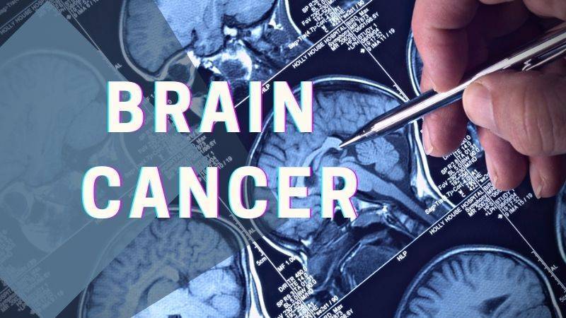
Histotripsy, a groundbreaking non-invasive ultrasonic technique, is rapidly gaining traction in the realm of cancer therapy. By harnessing the power of cavitation, histotripsy meticulously ablates targeted tissues, positioning itself as a potential successor to traditional cancer treatments. This comprehensive article seeks to unravel the intricate immunological responses instigated by histotripsy and its profound implications for cancer therapy.
What is the immunological response of Histotripsy?
At its core, histotripsy ablation is driven by a phenomenon known as cavitation. This intricate process involves the meticulous generation of microbubbles within the targeted tissues, culminating in their systematic breakdown. As these tissues undergo ablation, cells are fragmented into subcellular components and acellular debris. The immunological aftermath of histotripsy, irrespective of its variant – mHIFU, BH, or CCH, remains consistent, primarily due to the analogous effects they exert on the targeted tissues. While potential nuances between these sub-therapies exist, current research has yet to pinpoint significant disparities in their immunologic outcomes.
How does Histotripsy decrease pro-tumor immune cells?
The tumor microenvironment is a complex ecosystem comprising various cell types, signaling molecules, and extracellular matrix components. Within this intricate network, certain immune cells, often termed as pro-tumor immune cells, play a pivotal role in promoting tumor growth and metastasis. These cells, which include tumor-associated macrophages (TAMs), myeloid-derived suppressor cells (MDSCs), and regulatory T cells (Tregs), actively suppress anti-tumor immune responses, thereby facilitating tumor progression.
a. Tumor-Associated Macrophages (TAMs):
TAMs are a subset of macrophages that are recruited to the tumor site and are educated by the tumor microenvironment to adopt a pro-tumor phenotype. These cells can promote tumor growth, angiogenesis, and metastasis while suppressing anti-tumor immune responses. Histotripsy’s ability to target and modulate TAMs is of significant interest. By disrupting the tumor microenvironment, histotripsy can potentially reprogram TAMs from a pro-tumor M2 phenotype to an anti-tumor M1 phenotype, thereby reversing their tumor-promoting effects.
b. Myeloid-Derived Suppressor Cells (MDSCs):
MDSCs are a heterogeneous group of immature myeloid cells that expand during cancer, inflammation, and infection. In the context of cancer, MDSCs play a crucial role in suppressing T cell responses and promoting tumor growth. Preliminary studies suggest that histotripsy may reduce the number and suppressive function of MDSCs in the tumor microenvironment, thereby enhancing anti-tumor immunity.
c. Regulatory T cells (Tregs):
Tregs are a subset of CD4+ T cells that play a critical role in maintaining immune homeostasis by suppressing excessive immune responses. However, in the tumor microenvironment, Tregs can inhibit anti-tumor immune responses, thereby facilitating tumor growth. The impact of histotripsy on Tregs is an area of active research. By disrupting the tumor microenvironment and releasing tumor antigens, histotripsy may modulate the function and recruitment of Tregs, potentially enhancing anti-tumor immunity.
d. The Interplay with Other Immune Cells:
Apart from the aforementioned pro-tumor immune cells, the tumor microenvironment also contains other immune cells like dendritic cells, natural killer cells, and B cells. The interplay between these cells and pro-tumor immune cells is complex and multifaceted. Histotripsy’s ability to modulate this intricate network holds immense therapeutic potential. For instance, by targeting pro-tumor immune cells, histotripsy may enhance the antigen-presenting function of dendritic cells, leading to a more robust activation of T cells and a potent anti-tumor immune response.
The tumor microenvironment is a dynamic and complex landscape where various immune cells interact and influence tumor progression. Histotripsy, with its unique mechanism of action, holds the potential to modulate this environment, targeting pro-tumor immune cells, and enhancing anti-tumor immunity. As research in this area continues to evolve, a deeper understanding of histotripsy’s impact on the tumor microenvironment will pave the way for more effective and targeted cancer therapies.
What is the role of Damage Associated Molecular Patterns (DAMPs)?
Damage Associated Molecular Patterns (DAMPs) are a group of intracellular molecules that have garnered significant attention in the realm of immunology and cellular biology. These molecules, under normal circumstances, reside within the confines of the cell and perform essential functions. However, when cells undergo stress, trauma, or damage, DAMPs are released into the extracellular environment, where they assume a new and critical role.
One of the primary functions of DAMPs upon their release is to signal the immune system of potential threats. They act as distress signals, alerting the body to the presence of damaged or dying cells. This is particularly evident in procedures like histotripsy, a non-invasive technique that mechanically disrupts tissue structures. During histotripsy, the cellular damage leads to the liberation of various DAMPs, each with its unique role in the ensuing immune response.
For instance, Calreticulin (CRT) is not just a mere bystander in this process. When released, CRT moves to the cell surface, where it aids in antigen presentation. This action effectively marks the damaged cells for elimination, kickstarting an adaptive immune response tailored to address the specific threat.
High Mobility Group is another pivotal DAMP. As a nuclear protein, its primary role within the cell is to stabilize DNA structures. However, once outside, HMGB1 becomes a potent pro-inflammatory agent. It binds to receptors on immune cells, amplifying the inflammatory response, which is crucial in situations where rapid immune action is needed.
Adenosine Triphosphate (ATP), commonly recognized as the primary energy currency of the cell, also plays a role in this immune signaling process. When found outside the cell, ATP acts as a danger signal. Immune cells, recognizing the abnormal presence of extracellular ATP, are prompted to migrate to the site of injury, further intensifying the immune response.
Heat Shock Proteins (HSPs) complete the ensemble of key DAMPs released during histotripsy. These proteins, typically produced in response to cellular stress, have a dual role. Inside the cell, they ensure the proper folding of proteins. However, when released, HSPs actively stimulate the immune system, further emphasizing their importance in the body’s defense mechanisms.
The intricate connection between DAMPs and the immune response holds profound implications, especially in the field of oncology. Tumors, by their very nature, suppress the immune system, allowing them to grow unchecked. However, the inflammation induced by DAMPs can be harnessed and directed against tumor cells. By combining histotripsy with other therapeutic strategies, such as immunotherapy, there’s an exciting potential to boost the body’s natural defenses against cancer. This synergy could revolutionize cancer treatments, offering hope to countless patients worldwide.
However, the consistency and spectrum of DAMP release post-histotripsy remain subjects of intense research. Variables such as tumor heterogeneity, nuances in histotripsy modalities, and the ablation’s magnitude could influence DAMP dynamics.
What is the role of Pro-Inflammatory Cytokines and Chemokines?
Cytokines and chemokines, molecular orchestrators of immune modulation, play pivotal roles post-damage. IFNγ, a renowned inflammatory mediator, has been identified across a plethora of therapies and tumor typologies. While it is instrumental in galvanizing anti-tumor cells and inducing tumor cell apoptosis, it can paradoxically also foster immune evasion in select tumors.
Other cytokines, including IL-6, IL-2, TNF, IL-8, IL-13, and IL-10, have also been spotlighted post-histotripsy. Their roles are multifaceted, oscillating between pro-inflammatory and tumor-promoting activities. Deciphering these molecular dances is paramount for harnessing histotripsy’s full therapeutic potential.
How does Histotripsy curtail cancer progression?
Histotripsy’s immunologic ripples extend to both the innate and adaptive arms of the immune system. In the aftermath of ablation, there’s a discernible surge in innate immune stalwarts like neutrophils, natural killer cells, dendritic cells, and macrophages. This influx is emblematic of a seismic shift towards a more immunostimulatory tumor milieu.
On the adaptive front, post-histotripsy landscapes have witnessed burgeoning populations of T helper cells and B cells. Their augmented presence is emblematic of a fortified immune response, potentially pivotal in curtailing disease progression.
How does Histotripsy engage the Adaptive Immune System?
For a holistic, systemic anti-tumor immune response, the generation of pristine tumor antigens is of paramount importance. While direct investigations into histotripsy’s impact on antigen quality remain in their infancy, indirect evaluations offer tantalizing hints of its potential in driving efficacious antigen presentation. This capability could catalyze enhanced systemic immune responses, solidifying histotripsy’s position as a formidable tool in the oncologic arsenal.
In conclusion, Histotripsy, with its innovative approach to cancer therapy, is poised to revolutionize the therapeutic landscape. By leveraging cavitation to target neoplastic tissues and modulating the immune response at multiple levels, it holds immense promise. As the tapestry of research continues to expand, a more profound understanding of histotripsy’s immunological intricacies will undoubtedly pave the way for its optimized and widespread application in oncology.


