Richard Ramirez’s Battle with B-cell Lymphoma

Night Stalker-Richard Ramirez's cancer journey

Highlights

Richard Ramirez, also known as “The Night Stalker,” is a well-known name in the annals of American criminal history.

Born Ricardo Leyva Muñoz Ramirez on February 29, 1960, he was notorious for his crime spree in Los Angeles and San Francisco between 1984 and 1985.

His infamous activities culminated in multiple counts of murder, rape, burglary, and sodomy, which made headlines nationwide and triggered widespread fear.

But besides his criminal history, there is another aspect of his life that has garnered attention – his battle with cancer.

Ramirez’s Disease

In the years following his conviction and subsequent imprisonment, Ramirez’s health began to deteriorate.

In 2013, the infamous serial killer was diagnosed with B-cell lymphoma, a type of cancer that begins in the B-cells – a type of white blood cell that plays a crucial role in fighting infections by producing antibodies.

What is B-Cell Lymphoma?

B-Cell LymphomaB-cell lymphoma is a type of cancer that primarily affects the B-cells, a type of white blood cell that’s integral to the immune system.

B-cells are responsible for producing antibodies to combat infections, making them a crucial part of our body’s defense mechanism.

The term “B-cell lymphoma” actually refers to a group of cancers, as there are several different types that can develop in different kinds of B-cells.

These lymphomas can be broadly categorized into two types:

  • Hodgkin’s lymphomas
  • Non-Hodgkin’s lymphomas

with the latter being more common.

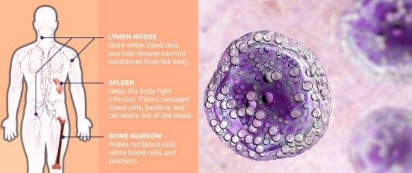
B-cell lymphomas can develop in various parts of the body, including the

  • Lymph nodes
  • Spleen
  • Bone marrow
  • Blood
  • Other organs where lymph tissue is found.

Types of B-Cell Lymphoma

Diffuse large B-cell Lymphoma

When your doctor diagnosis you with B-cell lymphoma, they’ll let you know exactly what kind you have.

The most common kind of non-Hodgkin’s lymphoma is known as Diffuse large B-cell lymphoma (DLBCL).

There are also other kinds of B-cell non-Hodgkin’s lymphoma, including:

  1. Follicular lymphoma: This type grows slowly and usually affects older people.

  2. Chronic lymphocytic leukemia/small lymphocytic leukemia (CLL/SLL): These are closely related types that grow slowly.

  3. Mantle cell lymphoma: This type grows quickly.

  4. Marginal zone lymphoma: This kind features small cells and grows slowly.

  5. Burkitt lymphoma: This is a rare disease that grows very fast.

  6. Lymphoplasmacytic lymphoma (Waldenstrom macroglobulinemia): This is a rare type that grows slowly.

  7. Primary mediastinal large B-cell lymphoma: This is a rare type that mostly affects young adults and is more common in women.

Warning Signs

Symptoms of B-Cell LymphomaThe symptoms of B-cell lymphoma can vary widely based on the specific type and the stage of the disease.

However, some common signs and symptoms often associated with this group of cancers include:

  1. Swollen lymph nodes: Often noticed in the neck, armpit, or groin, these are usually painless and one of the first signs of lymphoma. However, swollen lymph nodes can also result from infections and are not exclusively indicative of lymphoma.

  2. Fever: Persistent or recurrent fevers can occur as the body responds to cancerous cells.

  3. Night sweats: These can be severe enough to soak clothing and bedding.

  4. Unexplained weight loss: Significant, unintentional weight loss could be a sign of many types of cancer, including lymphoma.

  5. Fatigue: This could be a result of the body’s energy being redirected to fighting the cancerous cells, or it could be due to the cancer cells affecting the body’s normal metabolic processes.

  6. Coughing or shortness of breath: This can occur if the lymphoma affects lymph nodes in the chest, causing them to press on the windpipe (trachea).

  7. Pain or bloating in the abdomen: This can occur if the lymphoma affects lymph nodes in the abdomen or if the disease enlarges the spleen.

  8. Loss of appetite: People with lymphoma sometimes feel full after only a small amount of food, leading to a loss of appetite.

  9. Itchy skin or rash: This could be a symptom of lymphoma, although there are many other more common causes of these symptoms.

These symptoms can also be caused by many other conditions, most of which are far less serious than cancer.

However, anyone experiencing persistent or unexplained symptoms should consult with a healthcare provider to ensure an accurate diagnosis and appropriate treatment.

Causes and Risk factors

It appears to develop from genetic mutations in the B-cells. These mutations cause the B-cells to grow and divide out of control, forming a tumor.

The abnormal cells can also continue to live when normal cells would die. Over time, these abnormal B-cells accumulate and form a tumor, leading to lymphoma.

Certain factors can increase the risk of developing B-cell lymphoma:

  1. Age: The risk of B-cell lymphoma increases with age.

  2. Compromised Immune System: Those with weakened immune systems, such as people living with HIV/AIDS or those who take immune-suppressing medications (like those used following organ transplants), are more susceptible.

  3. Autoimmune Diseases: Conditions that cause the immune system to attack the body’s own cells, such as rheumatoid arthritis or lupus, can increase the risk of B-cell lymphoma.

  4. Exposure to Certain Chemicals: Certain chemicals, particularly those associated with specific industries like farming, can increase the risk. For example, exposure to pesticides or solvents has been linked to an increased risk of lymphomas.

  5. Infections: Certain viral and bacterial infections can increase the risk of specific types of non-Hodgkin’s lymphoma. Examples include the Epstein-Barr virus, which causes mononucleosis, the human T-cell leukemia/lymphoma virus (HTLV-1), Hepatitis C, and the bacterium Helicobacter pylori.

  6. Family History: Having a close relative who has had lymphoma can slightly increase the risk of the disease.

However, many people with these risk factors never develop lymphoma, and many who do get the disease have none of these risk factors.

As with many types of cancer, the development of B-cell lymphoma likely involves a complex interaction of genetic, environmental, and lifestyle factors.

Diagnosis

Diagnosis of B-Cell Lymphoma

Diagnosing B-cell lymphoma usually involves a series of steps to help confirm the presence of cancer cells, identify the type of lymphoma, and determine how far it’s spread (the stage).

Here’s a simplified process of how B-cell lymphoma is typically diagnosed:

  1. Medical History and Physical Examination: The process begins with a thorough review of the patient’s medical history and a physical exam. The doctor will ask about symptoms, lifestyle, past health issues, and family history of diseases. They will also check for physical signs of lymphoma such as swollen lymph nodes in the neck, underarms, or groin, as well as swelling or pain in the abdomen.

  2. Blood Tests: Blood tests can provide information about the overall health and how well the organs are functioning. They can also sometimes show signs of lymphoma.

  3. Biopsy: A biopsy is usually needed to definitively diagnose lymphoma. This involves taking a small sample of tissue from an enlarged lymph node or other affected area, which is then examined under a microscope by a pathologist. The pathologist looks for cancerous cells and can identify the specific type of lymphoma.

  4. Bone Marrow Examination: This test may be done to see if the lymphoma has spread to the bone marrow. A small sample of bone marrow is removed, usually from the hip bone, using a long, fine needle, and then examined under a microscope.

  5. Imaging Tests: Tests such as CT, PET, or MRI scans can help to determine the spread of the disease throughout the body. This helps in “staging” the disease, which in turn helps determine the best treatment options.

  6. Lumbar Puncture (Spinal Tap): This might be done if there’s a concern that the lymphoma has spread to the spinal fluid. A small sample of cerebrospinal fluid (CSF) is removed from the spinal canal and checked for lymphoma cells.

Lumbar Puncture(Spinal Tap)

Experiencing potential symptoms of lymphoma to seek medical advice as early as possible, as early detection can significantly improve the prognosis and treatment outcomes.

Treatment

The treatment for B-cell lymphoma can vary widely depending on the type of lymphoma, its stage, the patient’s overall health, and the patient’s personal preferences.

Here are some commonly used treatment options for B-cell lymphoma:

  1. Chemotherapy: This is often the first line of treatment for many types of lymphoma. It involves using drugs to kill cancer cells. These drugs can be given intravenously or orally and work by interrupting the cell division process of the cancer cells.

  2. Radiation therapy: This treatment uses high-energy beams, like X-rays or protons, to kill cancer cells. It’s often used for treating early-stage lymphomas or as a palliative treatment to relieve symptoms in advanced stages of the disease.

  3. Immunotherapy: This treatment harnesses the power of the patient’s immune system to fight cancer. Drugs like rituximab (Rituxan) are used in some B-cell lymphomas to enhance the body’s natural defenses against the cancer.

  4. Targeted therapy: These are drugs that specifically target the vulnerabilities of the cancer cells. For example, ibrutinib (Imbruvica) and idelalisib (Zydelig) block a specific protein that B-cell lymphomas need to survive and grow.

  5. CAR-T cell therapy: This is a relatively new type of treatment where a patient’s T-cells (a type of immune system cell) are genetically engineered to produce receptors on their surface called chimeric antigen receptors (CARs). These receptors can recognize and kill the cancer cells. This therapy is usually considered for patients whose cancer has not responded to other treatments.

  6. Stem cell transplant: This procedure allows doctors to use higher doses of chemotherapy than usual. After chemotherapy, the patient receives a transplant of stem cells to help rebuild their bone marrow.

  7. Radioimmunotherapy: This treatment combines a radioactive particle with an antibody that targets the cancer cells. The antibody delivers the radiation directly to the cancer cells, limiting the damage to healthy cells.

Each of these treatments has potential side effects, and the best approach often involves a combination of different treatments.

Living With

Living with B-cell lymphoma can present several challenges, as it requires not only dealing with the physical effects of the disease and its treatments but also managing the emotional and psychological impacts.

Here are some aspects to consider when living with B-cell lymphoma:

  1. Managing Symptoms and Side Effects: Both the disease itself and its treatments can cause various symptoms and side effects, such as fatigue, pain, nausea, and weight changes. It’s essential to communicate these issues with your healthcare team, as they can suggest ways to manage them.

  2. Maintaining Nutrition: Proper nutrition can help you feel better, maintain strength, and cope with the side effects of treatment. A dietitian can help create a dietary plan that meets your specific needs.

  3. Staying Active: While it’s important to rest and take care of your body, maintaining a level of physical activity can improve your mood, help manage side effects like fatigue, and generally promote better health.

  4. Psychological and Emotional Support: It’s normal to experience a range of emotions when living with lymphoma, including anxiety and depression. Speaking with a therapist or counselor, joining a support group, or confiding in close friends or family can help manage these feelings.

  5. Regular Monitoring and Follow-up: Regular check-ups are critical to monitor the progress of the disease, assess how well the body is responding to treatment, and make adjustments as necessary.

  6. Financial Considerations: The cost of cancer treatment can be a significant concern. Social workers or financial advisors familiar with healthcare situations can help explore what financial assistance, insurance coverage, or funding options may be available.

  7. Communication: Keep an open line of communication with your healthcare team. Don’t hesitate to ask questions about anything you don’t understand or worry about.

Ramirez’s Survival

Richard Ramirez passed away on June 7, 2013, at Marin General Hospital in Greenbrae, California, less than three months after his cancer diagnosis.

He was 53 years old. Given Ramirez’s imprisonment, he likely had limited personal support and faced additional challenges managing his health condition.

His journey was undoubtedly marked by the physical discomfort and emotional strain typical of cancer, but it was also uniquely framed by his status as a death row inmate.

While it is difficult to determine how long he may have been living with the disease before it was diagnosed, his case illustrates the often aggressive nature of B-cell lymphomas and the importance of early detection and treatment.

In a Nutshell

Ramirez’s experience with B-cell lymphoma underscores that cancer can affect anyone, regardless of their circumstances.

His journey with the disease, however, was marked by unique challenges given his status as a prisoner, limiting his access to support and potentially impacting his overall care and treatment experience.

Share:

Facebook
WhatsApp
Aviral
Reviewed By : Dr. Aviral Vatsa

Social Media

Most Popular

Get The Latest Updates

Subscribe To Our Weekly Newsletter

No spam, notifications only about new products, updates.

Categories

On Key

Related Posts

Non-Invasive Treatment of Breast Cancer Using Histotripsy

Breast cancer is a global health challenge, with its impact felt across all corners of the world. According to the World Health Organization (WHO), breast cancer is the most frequent cancer among women, impacting 2.1 million women each year. It causes the greatest number of cancer-related deaths among women. In 2020, it is estimated that 685,000 women died from breast cancer worldwide. As the incidence of breast cancer rises, particularly in developing countries where the majority of cases are diagnosed in late stages, the need for accessible and non-invasive treatments is more pressing than ever. Histotripsy promises to be a gamechanger for the non-invasive treatment of breast cancer worldwide. In India, breast cancer has overtaken cervical cancer as the most common cancer among women. The Indian Council of Medical Research (ICMR) has reported a significant rise in cases, with breast cancer accounting for 14% of cancers in Indian women. Urban areas register a higher incidence rate than rural areas, with cities like Delhi, Mumbai, and Kolkata showing increasing numbers. Late diagnosis is a significant issue, with a majority of breast cancer patients presenting at stage 2 or later, which can limit treatment options and reduce survival rates. Globally, the advancements in histotripsy research are paving the way for its potential application in breast cancer treatment. The technology’s precision and the possibility of outpatient procedures could lead to a paradigm shift in cancer care, offering a treatment that is not only effective but also aligns with the needs and preferences of patients. The introduction of histotripsy could be particularly transformative in India, where the healthcare system is burdened by the high volume of patients and often limited resources. The non-invasive nature of histotripsy could provide a less resource-intensive treatment option, reducing the need for surgical facilities and lengthy hospital stays. This could be a game-changer for rural and underserved areas where access to surgical oncologists and specialized care is limited. For countries like India, where cultural factors often influence healthcare decisions, the ability to preserve the breast could lead to increased acceptance and adherence to treatment protocols. The Potential Impact of Histotripsy in Treating Breast Cancer In the context of breast cancer, histotripsy has been shown to effectively reduce tumor burden in both subcutaneous and orthotopic mouse models, indicating its potential for treating primary breast tumors. The immunological responses to histotripsy ablation are mediated by the release of tumor antigens and damage-associated molecular patterns (DAMPs) into the tumor microenvironment. These factors can potentiate the effects of checkpoint inhibition, even sensitizing previously resistant cancers to immunotherapy. Histotripsy has also been shown to promote local and systemic immunological responses, reducing tumor burden in breast cancer. This is achieved by lysing target tumor cells, which release tumor antigens and DAMPs into the tumor microenvironment. The immunomodulatory impact of histotripsy may be key to expanding the impact and promise of cancer immunotherapy. In addition, histotripsy has the potential to be used in combination with other cancer treatments, such as chemotherapy and immunotherapy, to enhance their effectiveness and reduce tumor burden. The immunomodulatory impact of histotripsy may also make it an attractive option for treating metastatic breast cancer, as it can promote the polarization of monocytes and macrophages to pro-anti-tumor phenotypes, reducing the magnitude of tumor-supporting immune cells within the tumor microenvironment. What Is Histotripsy? Histotripsy is a promising non-invasive, non-ionizing, non-thermal ablation modality for the treatment of breast cancer. It initiates local and systemic immunological responses, reduces tumor burden, and promotes an anti-tumor microenvironment. The immunomodulatory impact of histotripsy may be key to expanding the impact and promise of cancer immunotherapy, making it an attractive option for treating primary and metastatic breast cancer. The potential impact of histotripsy in treating breast cancer could be significant due to several key advantages: 1. Non-invasive Treatment Histotripsy’s non-invasive nature stands out as one of its most significant benefits. Traditional cancer surgeries involve incisions, which come with risks such as infection, longer hospital stays, and longer recovery periods. By using focused ultrasound waves, histotripsy can destroy cancerous tissues without making any cuts. This approach can reduce the overall stress on the patient’s body, potentially leading to quicker recovery and less physical trauma. It could be particularly advantageous for patients who are poor candidates for surgery due to other health issues. 2. Precision The accuracy of histotripsy is due to its ability to focus ultrasound waves very precisely on a target area, often with millimeter accuracy. This precision is crucial in the treatment of breast cancer, where the goal is to remove or destroy cancerous cells without damaging the surrounding healthy tissue. Such targeted therapy could lead to better preservation of breast tissue, which is important for the patient’s physical and psychological recovery, potentially improving cosmetic outcomes and reducing the need for reconstructive surgery. 3. Reduced Side Effects Conventional treatments like chemotherapy and radiation can have significant side effects because they are not able to distinguish between healthy and cancerous cells. Histotripsy’s targeted approach could minimize this issue, as the therapy focuses only on the cancerous tissues. This targeted disruption means the body may be less overwhelmed by toxins and the side effects of cell death, possibly resulting in a better quality of life during and after treatment. 4. Repeatable treatment One of the limitations of radiation therapy is that there is a maximum total dose that can be safely administered to a patient. This cap does not exist with histotripsy. Because histotripsy does not use ionizing radiation, treatments can be administered multiple times without the cumulative risks associated with radiation. This repeatability allows clinicians to adjust treatment plans based on how the cancer responds and could be especially beneficial for aggressive or recurring breast cancers. 5. Potential in Drug Delivery Research has suggested that the mechanical disruption of tumor tissues by histotripsy can increase the permeability of those tissues. This change could potentially enhance the delivery and efficacy of chemotherapy drugs by allowing higher concentrations of the drug to penetrate the tumor. Furthermore, the combination of histotripsy and

Non-Invasive Treatment of Liver Tumors: The Revolutionary Role of Histotripsy

Liver tumors, whether benign or malignant, have long been a significant health concern. Traditional treatments, while effective, often involve invasive surgical procedures that come with inherent risks and complications. For many patients, especially those with small, numerous, or strategically located tumors, surgery might not be a viable option. However with Histotripsy, a groundbreaking procedure that promises a non-invasive alternative with remarkable potential, effective non-invasive treatment liver cancer is now possible. Histotripsy is a novel medical procedure that harnesses the power of ultrasound waves to mechanically disintegrate tissue structures. Unlike other treatments that depend on heat or radiation, histotripsy utilizes the sheer force of sound waves. These waves are meticulously focused on the target tissue, leading to its fragmentation without harming the surrounding healthy tissue. The precision and non-invasive nature of histotripsy make it a beacon of hope for liver tumor patients. A Closer Look at Evidence Research has demonstrated the efficacy of histotripsy in creating safe and effective ablations in the in vivo human-scale porcine normal liver. In various studies, target liver volumes ranging from 12–60 ml were completely ablated in durations spanning 20–75 min. The results were consistent and promising: within the ablated region, there was uniform tissue disruption with no viable cells remaining. Impressively, major vessels and bile ducts remained intact. The realm of medical treatments has always been complemented by the power of visual evidence, providing clinicians, researchers, and patients with tangible proof of a procedure’s effectiveness. Magnetic Resonance Imaging (MRI) stands at the forefront of this visual exploration. The high-resolution images produced by MRI scanners offer a detailed look into the internal structures of the liver, both before and after histotripsy treatment. Specifically, axial T2-weighted MR images have been invaluable (Figure 1). These images vividly depict the ablation volume, which is the targeted area that underwent histotripsy. The clarity and precision of these images allow medical professionals to ascertain the exact boundaries of the treated area, ensuring that the tumor cells have been effectively disrupted while leaving the surrounding healthy liver tissues untouched. Complementing the MRI scans are gross morphological examinations. These examinations, often conducted post-procedure, provide a hands-on, macroscopic view of the liver. They reveal a consistent pattern of tissue disruption within the ablation zone, characterized by a lack of viable hepatocytes, which are the primary cells of the liver. This uniformity in tissue disruption is a testament to histotripsy’s precision, ensuring that the treatment is both thorough and targeted. Furthermore, the visual evidence extends beyond just the immediate aftermath of the procedure. MR images from rodent Hepatocellular Carcinoma (HCC) models, a common type of liver cancer, provide insights into the longer-term effects of histotripsy. Initial scans of these models show tumors with a hyperintense signal on T2-weighted MRI, indicating active and aggressive tumor growth. However, post-histotripsy images paint a different picture. The previously hyperintense tumors now display a T2 hypointense signal within the ablation zone, signifying successful disruption of the tumor cells. Even more promising are follow-up scans taken 12 weeks after the procedure. These images often reveal a near-total disappearance of the tumor, replaced by a small fibrous tissue zone. This transformation underscores histotripsy’s potential not just as a treatment method but as a possible path to long-term recovery. The visual evidence supporting histotripsy’s effectiveness is both compelling and comprehensive. From high-resolution MRI scans to hands-on morphological examinations, each piece of evidence builds a case for histotripsy as a revolutionary, non-invasive treatment for liver tumors. What are the benefits of Histotripsy for Liver Tumor Treatment? The Immune Response of Histotripsy Another fascinating aspect of histotripsy is its potential to induce an immune response. FACS analysis of histotripsy-ablated tumors identified significant levels of intratumoral CD8+ T cell infiltration, much higher than untreated control tumors. This suggests that histotripsy might play a role in boosting the body’s natural defenses against cancer cells. Additionally, there was a noticeable reduction in pulmonary metastases in mice treated with histotripsy compared to untreated controls. Given that bones, especially ribs, are highly reflective and absorptive for ultrasound propagation, the feasibility and safety of histotripsy through ribs were meticulously studied. The results were reassuring. Ablation zones created through full ribcage coverage were comparable to those with only overlying soft tissue. The temperature increase to ribs was minimal, ensuring no thermal damage to the ribs or surrounding tissue. However, it’s worth noting that in one paper by Smolock et al., body wall damage was reported. This was likely due to pre-focal cavitation on the ribs. But subsequent studies addressed this by adjusting the focal pressure and duty cycle, effectively eliminating such damage. In some cases, transient thrombosis in portal and hepatic veins was observed within the treatment zone, akin to outcomes from radiofrequency and microwave ablation. The long-term response to liver treatment by histotripsy has also been studied. In normal rodent models, the acellular homogenate generated by histotripsy was absorbed within a month, leaving only a minuscule fibrous region. In tumor treatment studies, tumors were completely absorbed within 7–10 weeks post-histotripsy, with no evidence of residual tumor after three months. In conclusion, Histotripsy is undeniably a game-changer in the realm of liver tumor treatments. Its non-invasive nature, combined with its precision and efficacy, makes it a promising alternative to traditional surgical interventions. As research continues and technology advances, histotripsy could very well become the gold standard for treating not just liver tumors but a plethora of other medical conditions. For patients and medical professionals alike, it heralds a future of safer, more effective, and less invasive treatment options.

Immunological Effects Of Histotripsy for Cancer Therapy

Histotripsy, a groundbreaking non-invasive ultrasonic technique, is rapidly gaining traction in the realm of cancer therapy. By harnessing the power of cavitation, histotripsy meticulously ablates targeted tissues, positioning itself as a potential successor to traditional cancer treatments. This comprehensive article seeks to unravel the intricate immunological responses instigated by histotripsy and its profound implications for cancer therapy. What is the immunological response of Histotripsy? At its core, histotripsy ablation is driven by a phenomenon known as cavitation. This intricate process involves the meticulous generation of microbubbles within the targeted tissues, culminating in their systematic breakdown. As these tissues undergo ablation, cells are fragmented into subcellular components and acellular debris. The immunological aftermath of histotripsy, irrespective of its variant – mHIFU, BH, or CCH, remains consistent, primarily due to the analogous effects they exert on the targeted tissues. While potential nuances between these sub-therapies exist, current research has yet to pinpoint significant disparities in their immunologic outcomes. How does Histotripsy decrease pro-tumor immune cells? The tumor microenvironment is a complex ecosystem comprising various cell types, signaling molecules, and extracellular matrix components. Within this intricate network, certain immune cells, often termed as pro-tumor immune cells, play a pivotal role in promoting tumor growth and metastasis. These cells, which include tumor-associated macrophages (TAMs), myeloid-derived suppressor cells (MDSCs), and regulatory T cells (Tregs), actively suppress anti-tumor immune responses, thereby facilitating tumor progression. a. Tumor-Associated Macrophages (TAMs): TAMs are a subset of macrophages that are recruited to the tumor site and are educated by the tumor microenvironment to adopt a pro-tumor phenotype. These cells can promote tumor growth, angiogenesis, and metastasis while suppressing anti-tumor immune responses. Histotripsy’s ability to target and modulate TAMs is of significant interest. By disrupting the tumor microenvironment, histotripsy can potentially reprogram TAMs from a pro-tumor M2 phenotype to an anti-tumor M1 phenotype, thereby reversing their tumor-promoting effects. b. Myeloid-Derived Suppressor Cells (MDSCs): MDSCs are a heterogeneous group of immature myeloid cells that expand during cancer, inflammation, and infection. In the context of cancer, MDSCs play a crucial role in suppressing T cell responses and promoting tumor growth. Preliminary studies suggest that histotripsy may reduce the number and suppressive function of MDSCs in the tumor microenvironment, thereby enhancing anti-tumor immunity. c. Regulatory T cells (Tregs): Tregs are a subset of CD4+ T cells that play a critical role in maintaining immune homeostasis by suppressing excessive immune responses. However, in the tumor microenvironment, Tregs can inhibit anti-tumor immune responses, thereby facilitating tumor growth. The impact of histotripsy on Tregs is an area of active research. By disrupting the tumor microenvironment and releasing tumor antigens, histotripsy may modulate the function and recruitment of Tregs, potentially enhancing anti-tumor immunity. d. The Interplay with Other Immune Cells: Apart from the aforementioned pro-tumor immune cells, the tumor microenvironment also contains other immune cells like dendritic cells, natural killer cells, and B cells. The interplay between these cells and pro-tumor immune cells is complex and multifaceted. Histotripsy’s ability to modulate this intricate network holds immense therapeutic potential. For instance, by targeting pro-tumor immune cells, histotripsy may enhance the antigen-presenting function of dendritic cells, leading to a more robust activation of T cells and a potent anti-tumor immune response. The tumor microenvironment is a dynamic and complex landscape where various immune cells interact and influence tumor progression. Histotripsy, with its unique mechanism of action, holds the potential to modulate this environment, targeting pro-tumor immune cells, and enhancing anti-tumor immunity. As research in this area continues to evolve, a deeper understanding of histotripsy’s impact on the tumor microenvironment will pave the way for more effective and targeted cancer therapies. What is the role of Damage Associated Molecular Patterns (DAMPs)? Damage Associated Molecular Patterns (DAMPs) are a group of intracellular molecules that have garnered significant attention in the realm of immunology and cellular biology. These molecules, under normal circumstances, reside within the confines of the cell and perform essential functions. However, when cells undergo stress, trauma, or damage, DAMPs are released into the extracellular environment, where they assume a new and critical role. One of the primary functions of DAMPs upon their release is to signal the immune system of potential threats. They act as distress signals, alerting the body to the presence of damaged or dying cells. This is particularly evident in procedures like histotripsy, a non-invasive technique that mechanically disrupts tissue structures. During histotripsy, the cellular damage leads to the liberation of various DAMPs, each with its unique role in the ensuing immune response. For instance, Calreticulin (CRT) is not just a mere bystander in this process. When released, CRT moves to the cell surface, where it aids in antigen presentation. This action effectively marks the damaged cells for elimination, kickstarting an adaptive immune response tailored to address the specific threat.  High Mobility Group  is another pivotal DAMP. As a nuclear protein, its primary role within the cell is to stabilize DNA structures. However, once outside, HMGB1 becomes a potent pro-inflammatory agent. It binds to receptors on immune cells, amplifying the inflammatory response, which is crucial in situations where rapid immune action is needed. Adenosine Triphosphate (ATP), commonly recognized as the primary energy currency of the cell, also plays a role in this immune signaling process. When found outside the cell, ATP acts as a danger signal. Immune cells, recognizing the abnormal presence of extracellular ATP, are prompted to migrate to the site of injury, further intensifying the immune response. Heat Shock Proteins (HSPs) complete the ensemble of key DAMPs released during histotripsy. These proteins, typically produced in response to cellular stress, have a dual role. Inside the cell, they ensure the proper folding of proteins. However, when released, HSPs actively stimulate the immune system, further emphasizing their importance in the body’s defense mechanisms. The intricate connection between DAMPs and the immune response holds profound implications, especially in the field of oncology. Tumors, by their very nature, suppress the immune system, allowing them to grow unchecked. However, the inflammation induced by DAMPs can be harnessed and directed

Histotripsy: A Revolution in Precise Tissue Ablation

Histotripsy has emerged as a beacon of innovation in the ever-evolving landscape of medical technology. It  promises a future where precise tissue ablation can be achieved without the invasiveness of traditional surgical methods. But what makes histotripsy stand out? The answer lies in its unique mechanism of action, which harnesses the power of ultrasound to mechanically disrupt tissue structures. This article delves deep into the mechanism of histotripsy and how it paves the way for precise tissue ablation, especially in the realm of cancer treatment. What is Histotripsy? Histotripsy, a groundbreaking medical technique, is rapidly gaining traction in the healthcare sector due to its potential to revolutionise tissue ablation procedures. The term “histotripsy” is derived from the Greek words “histo,” meaning tissue, and “tripsy,” meaning to break. Histotripsy is a non-invasive approach to tissue disruption, and its unique mechanism of action  sets it apart from any other innovations. . The concept of histotripsy was first introduced at the University of Michigan in 2004. Since its inception, the technique has undergone significant advancements, with researchers continually exploring its potential applications and refining its methodology. The term itself encapsulates the essence of the procedure: a method to break down soft tissue. How does Histotripsy work? The Fundamental Mechanism: Cavitation  Cavitation, a phenomenon central to histotripsy, refers to the formation, growth, and subsequent collapse of gas or vapour-filled bubbles within a liquid medium when subjected to rapid pressure changes. In the context of histotripsy, the human body’s tissue serves as this liquid medium, and high-intensity, short-duration ultrasound pulses induce the rapid pressure changes. When tissues are exposed to these potent ultrasound pulses, the alternating high and low pressures lead to the creation of minuscule bubbles or cavities. These bubbles might initially form around pre-existing gas pockets or microscopic impurities within the tissue. As the ultrasound pulses persist, these bubbles expand due to the negative pressure phases of the ultrasound wave. Bubble Dynamics: The Heart of Tissue Disruption  The true essence of histotripsy is realised during the bubble collapse phase. Following the negative phase, the positive pressure phase of the ultrasound wave causes the expanded bubbles to undergo a swift and violent implosion. This rapid collapse generates potent local shock waves and produces high-velocity liquid jets. These intense mechanical forces, stemming from both the shock waves and the jets, act upon the surrounding tissue. The outcome is a mechanical breakdown of the tissue at a cellular level, resulting in the tissue being fractionated into a liquefied form. This liquid consists of a homogenised blend of cell debris and the extracellular matrix. How does Histotripsy achieve precision in action? In the realm of medical interventions, precision is paramount. The ability to target specific tissues or cells without affecting the surrounding structures can be the difference between successful treatment and unintended complications. Histotripsy, with its groundbreaking approach to tissue ablation, exemplifies this principle of precision in action. Let’s delve deeper into how histotripsy achieves such unparalleled accuracy. Histotripsy employs high-intensity ultrasound pulses to induce cavitation within the targeted tissue. The beauty of this technique lies in the ability to focus these ultrasound beams to a specific point, known as the focal zone. Within this focal zone, the energy of the ultrasound waves is concentrated, ensuring that the cavitation-induced tissue disruption occurs primarily within this localised area. This means that only the tissue within the focal zone is affected, while the surrounding structures remain untouched. One of the standout features that bolster histotripsy’s precision is the integration of real-time imaging. As the ultrasound waves are administered, they not only induce cavitation but also provide a live visual feed of the treatment area. This dual capability allows clinicians to monitor the formation and collapse of bubbles in real-time. Such immediate feedback ensures that the treatment is progressing as intended and allows for on-the-fly adjustments. If, for instance, the bubbles are not forming in the desired location or pattern, the clinician can instantly modify the parameters to achieve the desired effect. The precision of histotripsy can be likened to the accuracy of a surgeon’s scalpel, but without the invasiveness of a blade. The controlled generation and collapse of microbubbles ensure that only the targeted cells or tissues are disrupted. This selectivity is especially crucial when treating tumours or lesions located close to vital organs or critical structures. For example, when targeting a tumour adjacent to a major blood vessel, the precision of histotripsy ensures that the vessel remains unharmed, reducing the risk of bleeding or other complications. In many medical treatments, especially those involving radiation or surgery, there’s always a concern about collateral damage to healthy tissues. Histotripsy’s precision minimises this risk. By confining the tissue disruption to the focal zone, histotripsy ensures that the surrounding healthy tissues are spared. This not only enhances the safety profile of the treatment but also promotes faster healing and recovery. What sets Histotripsy apart from other cancer treatments? Histotripsy’s distinctive non-thermal approach to tissue ablation offers a fresh perspective in the realm of medical interventions. While many therapeutic ultrasound techniques, such as High-Intensity Focused Ultrasound (HIFU), rely on generating heat to achieve therapeutic effects, histotripsy stands apart. Traditional methods work by raising the temperature of the targeted tissue to a point where cellular proteins denature, leading to cell death. Although effective, this thermal approach has inherent risks. Elevated temperatures can inadvertently damage surrounding healthy tissues, especially if the heat spreads beyond the targeted area. Moreover, tissues sensitive to heat, like neural tissues, can be at risk of unintended damage. In contrast, histotripsy operates on a fundamentally different principle. Instead of using heat, it employs mechanical forces to achieve tissue disruption. This is achieved through the controlled generation and violent collapse of microbubbles within the tissue, a process known as cavitation. The implosive collapse of these bubbles generates intense local shock waves and produces high-speed liquid jets. These forces act on the tissue, leading to mechanical breakdown at the cellular level without the need for heat. The non-thermal nature of histotripsy offers