What did David Bowie suffer from? What is cancer du foie?

cancer du foie (liver cancer)

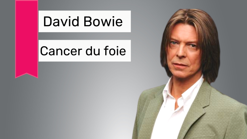
“Look up here – I’m in heaven,” sang David Bowie in “Lazarus,” a single from his last album Blackstar, which was released two days before his demise on January 10, 2016. 

cancer du foie (liver cancer)

Bowie had been suffering from cancer du foie, also referred to as liver cancer, for over 18 months. His last album, critics argue, seems to be a foreshadowing of his farewell to the world.

To understand Bowie’s unexpected journey with cancer du foie, let us get to know more about the disease.

What is cancer du foie?

Located on the right side of the lower ribs, the liver is the second largest organ of the human body. 

cancer du foie (liver cancer)

Its primary functions are as follows:

  • Breaks down and stores multiple nutrients: Some nutrients ought to be changed/metabolised by the liver in order to be utilised by the body for building energy or repairing body tissues. 
  • Conversion of fat into energy: It converts the food, particularly carbohydrates and fat, that you consume into energy through chemicals.
  • Makes Bile: The liver produces bile, a chemical that aids in digestion. Bile is stored in a small organ below the liver called the gallbladder and passes into the bowel through the bile duct.

cancer du foie (liver cancer) 

  • Makes protein: The liver is also responsible for producing proteins such as albumin. 
  • Aids in clotting: The liver produces substances which clot the blood and helps in healing injuries such as cuts. 
  • Breaks down toxic substances: It breaks down toxic substances, like alcohol, drugs etc., and rids them from your body through urination and excretion. 

When cancer du foie originates from the liver, it is referred to as primary liver cancer. Depending on the type of cells and location of origin, the type of primary liver cancer varies. 

Types of cancer du foie are: 

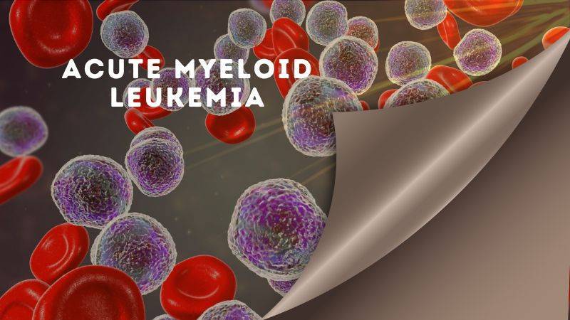
  • Hepatocellular carcinoma (HCC) is the most common type of liver cancer in adults. It has several subtypes. Fibrolamellar cancer is one such subtype which is typically rare. American Cancer Society estimates that it makes up less than 1% of HCCs.
  • Bile duct cancer, also referred to as Intrahepatic cholangiocarcinoma, originates from the bile ducts within or outside the liver. American Cancer Society states that “About 10% to 20% of cancers that start in the liver are intrahepatic cholangiocarcinomas.”
  • Angiosarcoma (or haemangiosarcoma) starts in the liver’s blood vessels and is extremely rare. It is considered harder to treat as the cancer cells spread faster in this type of cancer du foie. 
  • Hepatoblastoma is a rare childhood cancer which typically develops in children younger than 4. 

What are the symptoms of cancer du foie?

It is relatively harder to spot the symptoms of cancer du foie as they occur in the later stage. These symptoms also occur from various other diseases, but cancer du foie also remains a possibility. So, it is suggested that you visit the doctor for a check-up if you experience any of the symptoms mentioned below:

  • Unintentional weight loss and decrease in appetite
  • Jaundice or yellowing of the skin and whites of the eyes

cancer du foie (liver cancer)

  • Itching
  • Nausea and feeling sick
  • Swollen tummy (abdomen)
  • An enlarged liver which feels like a fullness under the ribs on the right side 

cancer du foie (liver cancer)

  • An enlarged spleen which feels like a fullness under the ribs on the left side 
  • Pain in the abdomen (belly) or near the right shoulder blade
  • Swelling or fluid build-up in the abdomen (belly)

What are the risk factors that lead to cancer du foie?

The development of cancer is dependent on various factors, such as genetics, age, lifestyle and so on. Thereby, these factors are classified as risk factors or possible causes that can lead to cancer du foie.  

They include:

  • Age: Although liver cancer is not limited to any age group, it is more common amongst older people. Cancer Research UK states that the highest rates are in 85 to 89-year-olds. Bowie was 68 when he was first diagnosed with cancer du foie. 
  • Liver cirrhosis: Cirrhosis is scarring of the liver due to previous damage which hinders the way the liver works and increases your chances of getting HCC or hepatocellular carcinoma. This can be caused by various factors. For instance, Bowie’s cancer was also linked with liver cirrhosis, which was caused by heavy drinking for a long period of time, unhealthy eating, non-alcoholic fatty liver disease, and hepatitis B and C.
  • Smoking: Cancer Research UK estimates that 20 out of 100 cases of liver cancer in the UK are caused by smoking. The case of Bowie was no different, as he began smoking at the age of 20. 

cancer du foie (liver cancer)

  • Body Weight and Diabetes: Obesity, unhealthy eating habits along with diabetes are also contributing factors in the development of cancer du foie. 
  • Alcohol: Long-term consumption of alcohol causes cirrhosis and can also damage the DNA inside the liver cell. According to Cancer Research UK, 7 out of 100 cases of liver cancer (7%) in the UK are caused by drinking alcohol.
  • Family history and previous health condition: Bowie’s previous health condition, particularly the six heart attacks that he survived, also contributed to the speedy multiplication of cancer cells.

Some other factors include:

  • Non-alcoholic fatty liver disease
  • Infection with hepatitis viruses
  • Human immunodeficiency virus (HIV) or AIDS
  • Aflatoxin
  • Betel quid
  • Gallstones or gallbladder removal
  • Chemicals
  • Liver flukes (parasitic worms)

Can you get cancer de foie? What are the chances of being diagnosed with cancer du foie?

According to Cancer Research UK, around 6,200 people are diagnosed with cancer du foie annually, estimated to be around 17 new cases each day in the UK.

Organisations such as the National Institute of Health (NIH) and the American Society of Clinical Oncology (ASCO) have analysed the prevalence of cancer du foie in the United States. According to SEER, approximately 1.1% of people in the US will be diagnosed with liver and intrahepatic bile duct cancer at some point during their lifetime, based on 2017–2019 data. 

ASCO further studies how the death rate of liver cancer doubled between 1980 and 2013. Cancer du foie is also labelled as the third leading cause of death globally. 

It is more common in men than in women. The risk of developing liver cancer gets higher as we get older.

According to COS, the statistics in France demonstrate that one in two men and one in three women will be diagnosed with cancer by age 85. 

How can cancer du foie be diagnosed?

Bowie was diagnosed with cancer du foie 18 months before his demise but had not publicised his condition. The average prognosis after a liver cancer diagnosis is generally about a year.

While liver cancer can be typically treated well after early diagnosis, advanced stage cancer is harder to treat, which must’ve been the case with Bowie. 

It can be detected earlier through regular screening via alpha-fetoprotein (AFP) blood tests and ultrasound exams, particularly in patients with a higher risk of liver cancer. 

This risk factor is determined by evaluating your previous medical history and physical examination. 

The diagnosis of cancer du foie can be detected through tests such as:

  • Imaging Tests

They use x-rays, magnetic fields, or sound waves to create pictures of the inside of your body. These include ultrasound, computed tomography (CT) scan, magnetic resonance imaging (MRI), angiography and bone scan. 

cancer du foie (liver cancer)

  • Biopsy

In a biopsy, the doctors take a tissue sample from a particular area and detect the cancer cells. This tissue is collected through various means such as needle, laparoscopic and surgical biopsy.

  • Lab Tests

These include varying blood tests. The test for AFP (Alpha-fetoprotein) in your blood is most commonly associated with cancer du foie. A high level of AFP can result from multiple factors. 

However, this test is helpful for people who are already diagnosed with liver cancer as it can trace the effectiveness of the treatment procedure. 

Other blood tests include viral hepatitis, blood clotting, complete blood count (CBC), kidney function, and liver function tests (LFTs).

 What are the stages of cancer du foie?

After the diagnosis, the doctors try to locate the spread of cancer cells which is referred to as the staging process. It is crucial in order to determine the best treatment. 

Cancer du foie ranges from stage I to stage IV; each successive stage signifies a higher range of cancer cells. This is also called the number staging system, where Stage I signifies the early stage and stage IV signifies the advanced stage. 

The stage of cancer du foie can be determined through various systems. The AJCC (American Joint Committee on Cancer) TNM system is typically used in the United States. It is based on three key pieces of information, the size of the tumour (T), the spread to nearby lymph nodes (N) and the spread (metastasis) to distant sites (M).  

Doctors also use the Barcelona Clinic Liver Cancer (BCLC) staging system and the Child-Pugh system. The latter is used to evaluate your liver’s performance, particularly in cirrhosis cases. On the other hand, BCLC is used to help make treatment decisions for hepatocellular carcinoma.

Some other systems to measure the stages of cancer du foie are the Cancer of the Liver Italian Program (CLIP) system and the Okuda system.

There is no particular staging system which is universal as each is used in different parts of the world and often serves distinct purposes. Therefore, they can’t be compared. To understand what staging system your doctor uses to determine the stage, be sure to question them about the same. 

What are the treatments available for cancer du foie?

Bowie began his treatment in mid-2014. But after a year, his cancer had metastasised and spread throughout his body. 

The treatment procedure is selected depending on various factors, such as:

  • The location of the cancer
  • The size/stage of the cancer
  • The type of cancer du foie
  • The age, general health, previous medical ailments, and condition of the liver

The treatment may include surgery, chemotherapy, using heat to destroy the cancer, using targeted medicines, and radiotherapy. The details of the procedure are as follows:

  • Surgery:

If the detected tumour is small in size, it is possible to remove it through a surgical operation. This is referred to as a partial hepatectomy, where only a part of the liver is removed. It is conducted on patients who don’t have severe cirrhosis.

It is even possible to conduct surgery if the growth of the cancer cells is limited to the organ, in which case a liver transplant would also be conducted. However, finding a donor is a long process, and patients with severe liver cirrhosis are considered unfit for a transplant. 

  • Chemotherapy:

This procedure eliminates cancer cells through the use of medicine and can be used both before and after the surgery. Before the surgery, it is used to shrink the size of the tumour to make it feasible for surgery. After the surgery, it is typically used to ensure that the cancer cells don’t return. 

The most common chemotherapy drugs for ministering liver cancer comprise of Gemcitabine (Gemzar), Oxaliplatin (Eloxatin), Cisplatin, Doxorubicin (pegylated liposomal doxorubicin), 5-fluorouracil (5-FU), Capecitabine (Xeloda), and Mitoxantrone (Novantrone). These drugs are often used in combination. 

Chemotherapy can be administered in different ways. In systemic chemotherapy, drugs are injected into the vein or taken orally. In regional chemotherapy, drugs are injected right into the vein that leads to the liver. Hepatic artery infusion (HAI), or chemo is given directly into the hepatic artery, is one such type.

cancer du foie (liver cancer)

HAI is slightly different from chemoembolisation because surgery is needed to put an infusion pump under the skin of the abdomen (belly). Embolisation infiltrates drugs directly into an artery in the liver to block or reduce the blood flow to a tumour in the liver. 

The liver has two blood supplies, the portal vein and the hepatic artery. The latter dominantly feeds cancer cells, so blocking it would shrink the possible growth of the tumour. 

  • Using heat to destroy the cancer:

Thermal ablation consists of the use of heat, which destroys the cancer cells without terminating them. It is used in cases where surgery is not a feasible solution. However, it is not as effective as surgery. 

Ablation is apt for tumours no larger than 3 cm across (slightly over an inch). It is sometimes used with embolisation for slightly larger tumours (1 to 2 inches or 3 to 5 cm across).

cancer du foie (liver cancer)

Some ablation techniques are radiofrequency ablation (RFA), microwave ablation (MWA), cryoablation (cryotherapy), and ethanol (alcohol) ablation. 

  • Radiation Therapy

During this procedure, the patient is subjected to high-level energy rays to kill the cancer cells. It is usually given through external beam radiation therapy (EBRT), which focuses radiation from a source outside of the body on the cancer cells. Newer radiation techniques are also emerging, such as stereotactic body radiation therapy (SBRT), which is shorter in duration. 

  • Targeted Drug Therapy

It works differently than standard chemotherapy drugs and has different side effects. While the standard chemotherapy eliminates cancer cells in general, this is opted to tackle liver cancer cells specifically. This includes drugs such as kinase inhibitors and monoclonal antibodies.

What is life like with cancer du foie?

Ivo Van Hove, who directed Bowie’s musical Lazarus, described Bowie as a “man fighting.” He says, “He fought like a lion and kept working like a lion through it all. He was fighting death, and he wanted to continue and continue. Afterwards, we were sitting behind the stage, and he said, ‘let’s start a second one now, the sequel to Lazarus’.”

cancer du foie (liver cancer)

For some people with cancer du foie, treatment can terminate or eliminate the cancer cells. The completion of the treatment can be both stressful and exciting.

Sometimes, the cancer cells get completely eradicated with minimal chances of returning. But for many people with cancer du foie, the cancer may never go away entirely, or it might recur in another part of the body. 

In the latter case, the patients may continue to receive regular treatments with chemotherapy, radiation therapy, or other therapies to monitor and limit the growth of the cancer cells and keep it under control for as much time as possible.

The follow-up care is crucial and consists of general questioning, exams and blood tests, such as alpha-fetoprotein (AFP), liver function tests (LFTs), imaging tests etc. 

It ranges from every 3 to 6 months for the first 2 years, then every 6 to 12 months and also evaluates any side effects that might’ve resulted from the treatment.

However, it is not just the physical health that remains the focus during the follow-up. Patients are also recommended to share and talk about their thoughts and feelings to help them cope. 

Bowie’s case fell into the latter to the point where he stopped his treatment altogether. Three months before Bowie’s death, he was working with music director Johan Renck who remarked, “He found out that it is over…. we’ll end treatment or whatever capacity that means that his illness has won.”

What are the survival rates of cancer du foie?

According to the NIH SEER report, the 5-Year relative survival rate is 20.8% from 2012 to 2018 in the United States. Relative survival estimates the percentage of patients expected to survive their cancer.

However, it is to keep in mind that the relative survival rate is based on a large group and is a general estimate that can’t be applied to individual cases as no two cases are entirely alike. 

The survival rate also depends on the stage of cancer du foie. If the spread of the cancer cells is limited to its place of origin, it is referred to as localised, which is broadly stage I. If the cancer has spread to other organs, it is called regional or distant. 

cancer du foie (liver cancer)The 5-year survival for cancer du foie is normally increased in younger people compared to older people. Cancer Research UK estimates that around 25% of people aged 15 to 39 survive cancer du foie, whereas the survival estimates decrease to 5% for people aged between 80 to 99. (borrowed from: https://seer.cancer.gov/statfacts/html/livibd.html)

What does the latest research on cancer du foie say?

The current research is globally oriented toward searching for better ways to prevent, diagnose and treat cancer du foie. Cancer Research UK documents various clinical trials which undertake this pursuit. 

For prevention, researchers are currently working toward developing a hepatitis C vaccine. For screening, researchers are trying to locate other biomarkers which may assist in a more accurate diagnosis of cancer du foie at an early stage.

Researchers are also drawing a comparative analysis of varying techniques to understand which is more effective. To reduce the risk of cancer cells returning, researchers are looking for an appropriate adjuvant treatment for cancer du foie. For instance, in one trial, researchers tested an immunotherapy drug called pembrolizumab to evaluate its effectiveness in the reduction of this risk.

Watch out this pace for more updates on new research.

Share:

Facebook
WhatsApp
Aviral
Reviewed By : Dr. Aviral Vatsa

Social Media

Most Popular

Get The Latest Updates

Subscribe To Our Weekly Newsletter

No spam, notifications only about new products, updates.

Categories

On Key

Related Posts

Non-Invasive Treatment of Breast Cancer Using Histotripsy

Breast cancer is a global health challenge, with its impact felt across all corners of the world. According to the World Health Organization (WHO), breast cancer is the most frequent cancer among women, impacting 2.1 million women each year. It causes the greatest number of cancer-related deaths among women. In 2020, it is estimated that 685,000 women died from breast cancer worldwide. As the incidence of breast cancer rises, particularly in developing countries where the majority of cases are diagnosed in late stages, the need for accessible and non-invasive treatments is more pressing than ever. Histotripsy promises to be a gamechanger for the non-invasive treatment of breast cancer worldwide. In India, breast cancer has overtaken cervical cancer as the most common cancer among women. The Indian Council of Medical Research (ICMR) has reported a significant rise in cases, with breast cancer accounting for 14% of cancers in Indian women. Urban areas register a higher incidence rate than rural areas, with cities like Delhi, Mumbai, and Kolkata showing increasing numbers. Late diagnosis is a significant issue, with a majority of breast cancer patients presenting at stage 2 or later, which can limit treatment options and reduce survival rates. Globally, the advancements in histotripsy research are paving the way for its potential application in breast cancer treatment. The technology’s precision and the possibility of outpatient procedures could lead to a paradigm shift in cancer care, offering a treatment that is not only effective but also aligns with the needs and preferences of patients. The introduction of histotripsy could be particularly transformative in India, where the healthcare system is burdened by the high volume of patients and often limited resources. The non-invasive nature of histotripsy could provide a less resource-intensive treatment option, reducing the need for surgical facilities and lengthy hospital stays. This could be a game-changer for rural and underserved areas where access to surgical oncologists and specialized care is limited. For countries like India, where cultural factors often influence healthcare decisions, the ability to preserve the breast could lead to increased acceptance and adherence to treatment protocols. The Potential Impact of Histotripsy in Treating Breast Cancer In the context of breast cancer, histotripsy has been shown to effectively reduce tumor burden in both subcutaneous and orthotopic mouse models, indicating its potential for treating primary breast tumors. The immunological responses to histotripsy ablation are mediated by the release of tumor antigens and damage-associated molecular patterns (DAMPs) into the tumor microenvironment. These factors can potentiate the effects of checkpoint inhibition, even sensitizing previously resistant cancers to immunotherapy. Histotripsy has also been shown to promote local and systemic immunological responses, reducing tumor burden in breast cancer. This is achieved by lysing target tumor cells, which release tumor antigens and DAMPs into the tumor microenvironment. The immunomodulatory impact of histotripsy may be key to expanding the impact and promise of cancer immunotherapy. In addition, histotripsy has the potential to be used in combination with other cancer treatments, such as chemotherapy and immunotherapy, to enhance their effectiveness and reduce tumor burden. The immunomodulatory impact of histotripsy may also make it an attractive option for treating metastatic breast cancer, as it can promote the polarization of monocytes and macrophages to pro-anti-tumor phenotypes, reducing the magnitude of tumor-supporting immune cells within the tumor microenvironment. What Is Histotripsy? Histotripsy is a promising non-invasive, non-ionizing, non-thermal ablation modality for the treatment of breast cancer. It initiates local and systemic immunological responses, reduces tumor burden, and promotes an anti-tumor microenvironment. The immunomodulatory impact of histotripsy may be key to expanding the impact and promise of cancer immunotherapy, making it an attractive option for treating primary and metastatic breast cancer. The potential impact of histotripsy in treating breast cancer could be significant due to several key advantages: 1. Non-invasive Treatment Histotripsy’s non-invasive nature stands out as one of its most significant benefits. Traditional cancer surgeries involve incisions, which come with risks such as infection, longer hospital stays, and longer recovery periods. By using focused ultrasound waves, histotripsy can destroy cancerous tissues without making any cuts. This approach can reduce the overall stress on the patient’s body, potentially leading to quicker recovery and less physical trauma. It could be particularly advantageous for patients who are poor candidates for surgery due to other health issues. 2. Precision The accuracy of histotripsy is due to its ability to focus ultrasound waves very precisely on a target area, often with millimeter accuracy. This precision is crucial in the treatment of breast cancer, where the goal is to remove or destroy cancerous cells without damaging the surrounding healthy tissue. Such targeted therapy could lead to better preservation of breast tissue, which is important for the patient’s physical and psychological recovery, potentially improving cosmetic outcomes and reducing the need for reconstructive surgery. 3. Reduced Side Effects Conventional treatments like chemotherapy and radiation can have significant side effects because they are not able to distinguish between healthy and cancerous cells. Histotripsy’s targeted approach could minimize this issue, as the therapy focuses only on the cancerous tissues. This targeted disruption means the body may be less overwhelmed by toxins and the side effects of cell death, possibly resulting in a better quality of life during and after treatment. 4. Repeatable treatment One of the limitations of radiation therapy is that there is a maximum total dose that can be safely administered to a patient. This cap does not exist with histotripsy. Because histotripsy does not use ionizing radiation, treatments can be administered multiple times without the cumulative risks associated with radiation. This repeatability allows clinicians to adjust treatment plans based on how the cancer responds and could be especially beneficial for aggressive or recurring breast cancers. 5. Potential in Drug Delivery Research has suggested that the mechanical disruption of tumor tissues by histotripsy can increase the permeability of those tissues. This change could potentially enhance the delivery and efficacy of chemotherapy drugs by allowing higher concentrations of the drug to penetrate the tumor. Furthermore, the combination of histotripsy and

Non-Invasive Treatment of Liver Tumors: The Revolutionary Role of Histotripsy

Liver tumors, whether benign or malignant, have long been a significant health concern. Traditional treatments, while effective, often involve invasive surgical procedures that come with inherent risks and complications. For many patients, especially those with small, numerous, or strategically located tumors, surgery might not be a viable option. However with Histotripsy, a groundbreaking procedure that promises a non-invasive alternative with remarkable potential, effective non-invasive treatment liver cancer is now possible. Histotripsy is a novel medical procedure that harnesses the power of ultrasound waves to mechanically disintegrate tissue structures. Unlike other treatments that depend on heat or radiation, histotripsy utilizes the sheer force of sound waves. These waves are meticulously focused on the target tissue, leading to its fragmentation without harming the surrounding healthy tissue. The precision and non-invasive nature of histotripsy make it a beacon of hope for liver tumor patients. A Closer Look at Evidence Research has demonstrated the efficacy of histotripsy in creating safe and effective ablations in the in vivo human-scale porcine normal liver. In various studies, target liver volumes ranging from 12–60 ml were completely ablated in durations spanning 20–75 min. The results were consistent and promising: within the ablated region, there was uniform tissue disruption with no viable cells remaining. Impressively, major vessels and bile ducts remained intact. The realm of medical treatments has always been complemented by the power of visual evidence, providing clinicians, researchers, and patients with tangible proof of a procedure’s effectiveness. Magnetic Resonance Imaging (MRI) stands at the forefront of this visual exploration. The high-resolution images produced by MRI scanners offer a detailed look into the internal structures of the liver, both before and after histotripsy treatment. Specifically, axial T2-weighted MR images have been invaluable (Figure 1). These images vividly depict the ablation volume, which is the targeted area that underwent histotripsy. The clarity and precision of these images allow medical professionals to ascertain the exact boundaries of the treated area, ensuring that the tumor cells have been effectively disrupted while leaving the surrounding healthy liver tissues untouched. Complementing the MRI scans are gross morphological examinations. These examinations, often conducted post-procedure, provide a hands-on, macroscopic view of the liver. They reveal a consistent pattern of tissue disruption within the ablation zone, characterized by a lack of viable hepatocytes, which are the primary cells of the liver. This uniformity in tissue disruption is a testament to histotripsy’s precision, ensuring that the treatment is both thorough and targeted. Furthermore, the visual evidence extends beyond just the immediate aftermath of the procedure. MR images from rodent Hepatocellular Carcinoma (HCC) models, a common type of liver cancer, provide insights into the longer-term effects of histotripsy. Initial scans of these models show tumors with a hyperintense signal on T2-weighted MRI, indicating active and aggressive tumor growth. However, post-histotripsy images paint a different picture. The previously hyperintense tumors now display a T2 hypointense signal within the ablation zone, signifying successful disruption of the tumor cells. Even more promising are follow-up scans taken 12 weeks after the procedure. These images often reveal a near-total disappearance of the tumor, replaced by a small fibrous tissue zone. This transformation underscores histotripsy’s potential not just as a treatment method but as a possible path to long-term recovery. The visual evidence supporting histotripsy’s effectiveness is both compelling and comprehensive. From high-resolution MRI scans to hands-on morphological examinations, each piece of evidence builds a case for histotripsy as a revolutionary, non-invasive treatment for liver tumors. What are the benefits of Histotripsy for Liver Tumor Treatment? The Immune Response of Histotripsy Another fascinating aspect of histotripsy is its potential to induce an immune response. FACS analysis of histotripsy-ablated tumors identified significant levels of intratumoral CD8+ T cell infiltration, much higher than untreated control tumors. This suggests that histotripsy might play a role in boosting the body’s natural defenses against cancer cells. Additionally, there was a noticeable reduction in pulmonary metastases in mice treated with histotripsy compared to untreated controls. Given that bones, especially ribs, are highly reflective and absorptive for ultrasound propagation, the feasibility and safety of histotripsy through ribs were meticulously studied. The results were reassuring. Ablation zones created through full ribcage coverage were comparable to those with only overlying soft tissue. The temperature increase to ribs was minimal, ensuring no thermal damage to the ribs or surrounding tissue. However, it’s worth noting that in one paper by Smolock et al., body wall damage was reported. This was likely due to pre-focal cavitation on the ribs. But subsequent studies addressed this by adjusting the focal pressure and duty cycle, effectively eliminating such damage. In some cases, transient thrombosis in portal and hepatic veins was observed within the treatment zone, akin to outcomes from radiofrequency and microwave ablation. The long-term response to liver treatment by histotripsy has also been studied. In normal rodent models, the acellular homogenate generated by histotripsy was absorbed within a month, leaving only a minuscule fibrous region. In tumor treatment studies, tumors were completely absorbed within 7–10 weeks post-histotripsy, with no evidence of residual tumor after three months. In conclusion, Histotripsy is undeniably a game-changer in the realm of liver tumor treatments. Its non-invasive nature, combined with its precision and efficacy, makes it a promising alternative to traditional surgical interventions. As research continues and technology advances, histotripsy could very well become the gold standard for treating not just liver tumors but a plethora of other medical conditions. For patients and medical professionals alike, it heralds a future of safer, more effective, and less invasive treatment options.

Immunological Effects Of Histotripsy for Cancer Therapy

Histotripsy, a groundbreaking non-invasive ultrasonic technique, is rapidly gaining traction in the realm of cancer therapy. By harnessing the power of cavitation, histotripsy meticulously ablates targeted tissues, positioning itself as a potential successor to traditional cancer treatments. This comprehensive article seeks to unravel the intricate immunological responses instigated by histotripsy and its profound implications for cancer therapy. What is the immunological response of Histotripsy? At its core, histotripsy ablation is driven by a phenomenon known as cavitation. This intricate process involves the meticulous generation of microbubbles within the targeted tissues, culminating in their systematic breakdown. As these tissues undergo ablation, cells are fragmented into subcellular components and acellular debris. The immunological aftermath of histotripsy, irrespective of its variant – mHIFU, BH, or CCH, remains consistent, primarily due to the analogous effects they exert on the targeted tissues. While potential nuances between these sub-therapies exist, current research has yet to pinpoint significant disparities in their immunologic outcomes. How does Histotripsy decrease pro-tumor immune cells? The tumor microenvironment is a complex ecosystem comprising various cell types, signaling molecules, and extracellular matrix components. Within this intricate network, certain immune cells, often termed as pro-tumor immune cells, play a pivotal role in promoting tumor growth and metastasis. These cells, which include tumor-associated macrophages (TAMs), myeloid-derived suppressor cells (MDSCs), and regulatory T cells (Tregs), actively suppress anti-tumor immune responses, thereby facilitating tumor progression. a. Tumor-Associated Macrophages (TAMs): TAMs are a subset of macrophages that are recruited to the tumor site and are educated by the tumor microenvironment to adopt a pro-tumor phenotype. These cells can promote tumor growth, angiogenesis, and metastasis while suppressing anti-tumor immune responses. Histotripsy’s ability to target and modulate TAMs is of significant interest. By disrupting the tumor microenvironment, histotripsy can potentially reprogram TAMs from a pro-tumor M2 phenotype to an anti-tumor M1 phenotype, thereby reversing their tumor-promoting effects. b. Myeloid-Derived Suppressor Cells (MDSCs): MDSCs are a heterogeneous group of immature myeloid cells that expand during cancer, inflammation, and infection. In the context of cancer, MDSCs play a crucial role in suppressing T cell responses and promoting tumor growth. Preliminary studies suggest that histotripsy may reduce the number and suppressive function of MDSCs in the tumor microenvironment, thereby enhancing anti-tumor immunity. c. Regulatory T cells (Tregs): Tregs are a subset of CD4+ T cells that play a critical role in maintaining immune homeostasis by suppressing excessive immune responses. However, in the tumor microenvironment, Tregs can inhibit anti-tumor immune responses, thereby facilitating tumor growth. The impact of histotripsy on Tregs is an area of active research. By disrupting the tumor microenvironment and releasing tumor antigens, histotripsy may modulate the function and recruitment of Tregs, potentially enhancing anti-tumor immunity. d. The Interplay with Other Immune Cells: Apart from the aforementioned pro-tumor immune cells, the tumor microenvironment also contains other immune cells like dendritic cells, natural killer cells, and B cells. The interplay between these cells and pro-tumor immune cells is complex and multifaceted. Histotripsy’s ability to modulate this intricate network holds immense therapeutic potential. For instance, by targeting pro-tumor immune cells, histotripsy may enhance the antigen-presenting function of dendritic cells, leading to a more robust activation of T cells and a potent anti-tumor immune response. The tumor microenvironment is a dynamic and complex landscape where various immune cells interact and influence tumor progression. Histotripsy, with its unique mechanism of action, holds the potential to modulate this environment, targeting pro-tumor immune cells, and enhancing anti-tumor immunity. As research in this area continues to evolve, a deeper understanding of histotripsy’s impact on the tumor microenvironment will pave the way for more effective and targeted cancer therapies. What is the role of Damage Associated Molecular Patterns (DAMPs)? Damage Associated Molecular Patterns (DAMPs) are a group of intracellular molecules that have garnered significant attention in the realm of immunology and cellular biology. These molecules, under normal circumstances, reside within the confines of the cell and perform essential functions. However, when cells undergo stress, trauma, or damage, DAMPs are released into the extracellular environment, where they assume a new and critical role. One of the primary functions of DAMPs upon their release is to signal the immune system of potential threats. They act as distress signals, alerting the body to the presence of damaged or dying cells. This is particularly evident in procedures like histotripsy, a non-invasive technique that mechanically disrupts tissue structures. During histotripsy, the cellular damage leads to the liberation of various DAMPs, each with its unique role in the ensuing immune response. For instance, Calreticulin (CRT) is not just a mere bystander in this process. When released, CRT moves to the cell surface, where it aids in antigen presentation. This action effectively marks the damaged cells for elimination, kickstarting an adaptive immune response tailored to address the specific threat.  High Mobility Group  is another pivotal DAMP. As a nuclear protein, its primary role within the cell is to stabilize DNA structures. However, once outside, HMGB1 becomes a potent pro-inflammatory agent. It binds to receptors on immune cells, amplifying the inflammatory response, which is crucial in situations where rapid immune action is needed. Adenosine Triphosphate (ATP), commonly recognized as the primary energy currency of the cell, also plays a role in this immune signaling process. When found outside the cell, ATP acts as a danger signal. Immune cells, recognizing the abnormal presence of extracellular ATP, are prompted to migrate to the site of injury, further intensifying the immune response. Heat Shock Proteins (HSPs) complete the ensemble of key DAMPs released during histotripsy. These proteins, typically produced in response to cellular stress, have a dual role. Inside the cell, they ensure the proper folding of proteins. However, when released, HSPs actively stimulate the immune system, further emphasizing their importance in the body’s defense mechanisms. The intricate connection between DAMPs and the immune response holds profound implications, especially in the field of oncology. Tumors, by their very nature, suppress the immune system, allowing them to grow unchecked. However, the inflammation induced by DAMPs can be harnessed and directed

Histotripsy: A Revolution in Precise Tissue Ablation

Histotripsy has emerged as a beacon of innovation in the ever-evolving landscape of medical technology. It  promises a future where precise tissue ablation can be achieved without the invasiveness of traditional surgical methods. But what makes histotripsy stand out? The answer lies in its unique mechanism of action, which harnesses the power of ultrasound to mechanically disrupt tissue structures. This article delves deep into the mechanism of histotripsy and how it paves the way for precise tissue ablation, especially in the realm of cancer treatment. What is Histotripsy? Histotripsy, a groundbreaking medical technique, is rapidly gaining traction in the healthcare sector due to its potential to revolutionise tissue ablation procedures. The term “histotripsy” is derived from the Greek words “histo,” meaning tissue, and “tripsy,” meaning to break. Histotripsy is a non-invasive approach to tissue disruption, and its unique mechanism of action  sets it apart from any other innovations. . The concept of histotripsy was first introduced at the University of Michigan in 2004. Since its inception, the technique has undergone significant advancements, with researchers continually exploring its potential applications and refining its methodology. The term itself encapsulates the essence of the procedure: a method to break down soft tissue. How does Histotripsy work? The Fundamental Mechanism: Cavitation  Cavitation, a phenomenon central to histotripsy, refers to the formation, growth, and subsequent collapse of gas or vapour-filled bubbles within a liquid medium when subjected to rapid pressure changes. In the context of histotripsy, the human body’s tissue serves as this liquid medium, and high-intensity, short-duration ultrasound pulses induce the rapid pressure changes. When tissues are exposed to these potent ultrasound pulses, the alternating high and low pressures lead to the creation of minuscule bubbles or cavities. These bubbles might initially form around pre-existing gas pockets or microscopic impurities within the tissue. As the ultrasound pulses persist, these bubbles expand due to the negative pressure phases of the ultrasound wave. Bubble Dynamics: The Heart of Tissue Disruption  The true essence of histotripsy is realised during the bubble collapse phase. Following the negative phase, the positive pressure phase of the ultrasound wave causes the expanded bubbles to undergo a swift and violent implosion. This rapid collapse generates potent local shock waves and produces high-velocity liquid jets. These intense mechanical forces, stemming from both the shock waves and the jets, act upon the surrounding tissue. The outcome is a mechanical breakdown of the tissue at a cellular level, resulting in the tissue being fractionated into a liquefied form. This liquid consists of a homogenised blend of cell debris and the extracellular matrix. How does Histotripsy achieve precision in action? In the realm of medical interventions, precision is paramount. The ability to target specific tissues or cells without affecting the surrounding structures can be the difference between successful treatment and unintended complications. Histotripsy, with its groundbreaking approach to tissue ablation, exemplifies this principle of precision in action. Let’s delve deeper into how histotripsy achieves such unparalleled accuracy. Histotripsy employs high-intensity ultrasound pulses to induce cavitation within the targeted tissue. The beauty of this technique lies in the ability to focus these ultrasound beams to a specific point, known as the focal zone. Within this focal zone, the energy of the ultrasound waves is concentrated, ensuring that the cavitation-induced tissue disruption occurs primarily within this localised area. This means that only the tissue within the focal zone is affected, while the surrounding structures remain untouched. One of the standout features that bolster histotripsy’s precision is the integration of real-time imaging. As the ultrasound waves are administered, they not only induce cavitation but also provide a live visual feed of the treatment area. This dual capability allows clinicians to monitor the formation and collapse of bubbles in real-time. Such immediate feedback ensures that the treatment is progressing as intended and allows for on-the-fly adjustments. If, for instance, the bubbles are not forming in the desired location or pattern, the clinician can instantly modify the parameters to achieve the desired effect. The precision of histotripsy can be likened to the accuracy of a surgeon’s scalpel, but without the invasiveness of a blade. The controlled generation and collapse of microbubbles ensure that only the targeted cells or tissues are disrupted. This selectivity is especially crucial when treating tumours or lesions located close to vital organs or critical structures. For example, when targeting a tumour adjacent to a major blood vessel, the precision of histotripsy ensures that the vessel remains unharmed, reducing the risk of bleeding or other complications. In many medical treatments, especially those involving radiation or surgery, there’s always a concern about collateral damage to healthy tissues. Histotripsy’s precision minimises this risk. By confining the tissue disruption to the focal zone, histotripsy ensures that the surrounding healthy tissues are spared. This not only enhances the safety profile of the treatment but also promotes faster healing and recovery. What sets Histotripsy apart from other cancer treatments? Histotripsy’s distinctive non-thermal approach to tissue ablation offers a fresh perspective in the realm of medical interventions. While many therapeutic ultrasound techniques, such as High-Intensity Focused Ultrasound (HIFU), rely on generating heat to achieve therapeutic effects, histotripsy stands apart. Traditional methods work by raising the temperature of the targeted tissue to a point where cellular proteins denature, leading to cell death. Although effective, this thermal approach has inherent risks. Elevated temperatures can inadvertently damage surrounding healthy tissues, especially if the heat spreads beyond the targeted area. Moreover, tissues sensitive to heat, like neural tissues, can be at risk of unintended damage. In contrast, histotripsy operates on a fundamentally different principle. Instead of using heat, it employs mechanical forces to achieve tissue disruption. This is achieved through the controlled generation and violent collapse of microbubbles within the tissue, a process known as cavitation. The implosive collapse of these bubbles generates intense local shock waves and produces high-speed liquid jets. These forces act on the tissue, leading to mechanical breakdown at the cellular level without the need for heat. The non-thermal nature of histotripsy offers